. 2009 Jun;27(6):1366-75.
doi: 10.1002/stem.50.

Matrix metalloproteinase 1 is necessary for the migration of human bone marrow-derived mesenchymal stem cells toward human glioma

Affiliations
Free PMC article

Matrix metalloproteinase 1 is necessary for the migration of human bone marrow-derived mesenchymal stem cells toward human glioma

Ivy A W Ho et al. Stem Cells. 2009 Jun.
Free PMC article

Abstract

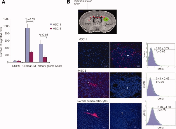
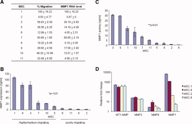
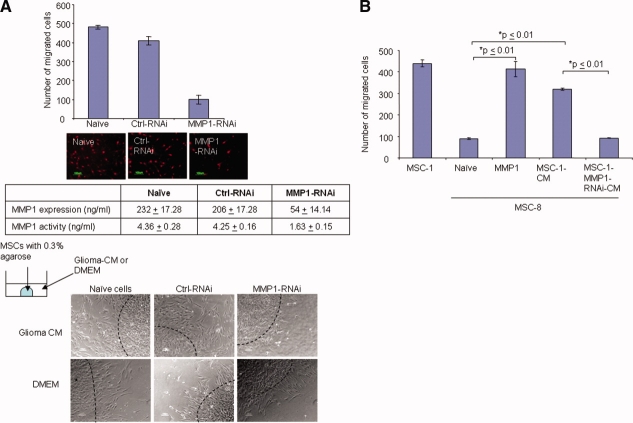
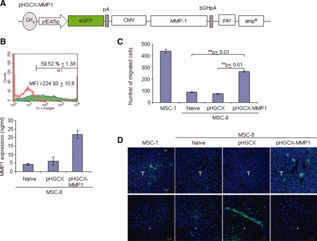
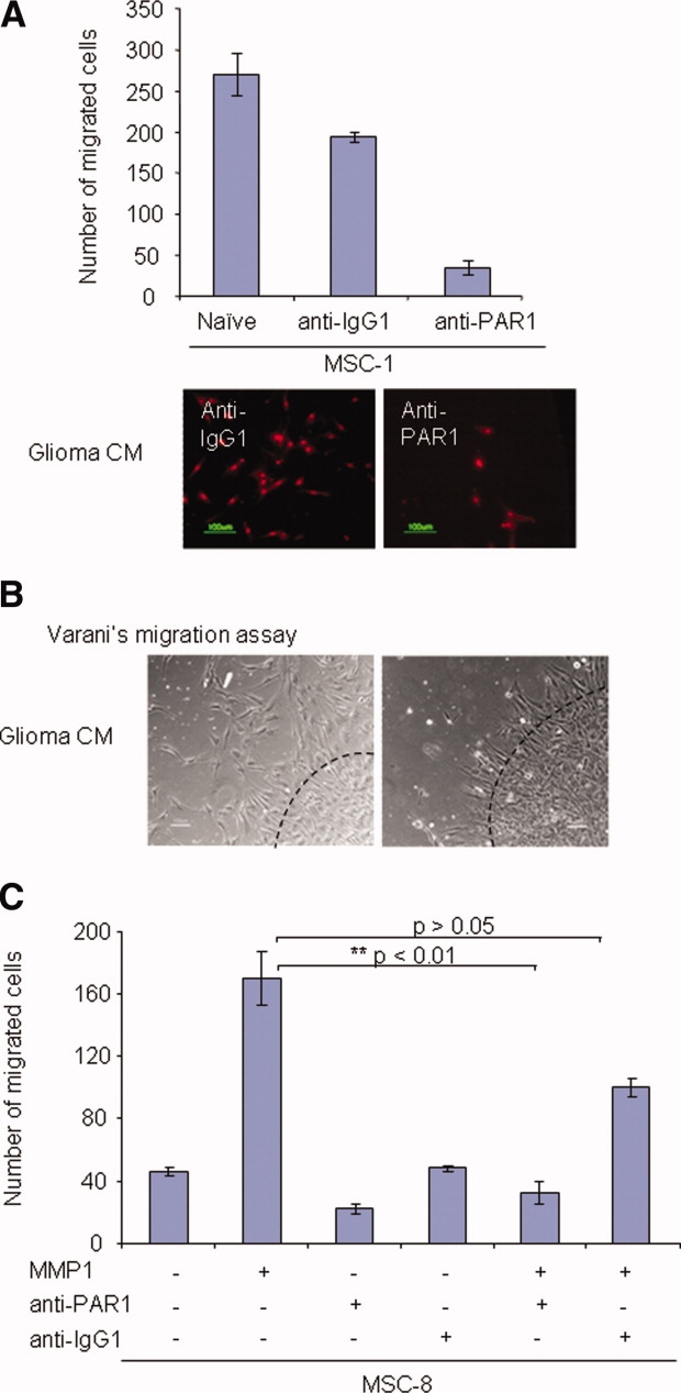
Human mesenchymal stem cells (MSCs) have increasingly been used as cellular vectors for the delivery of therapeutic genes to tumors. However, the precise mechanism of mobilization remains poorly defined. In this study, MSCs that expressed similar cell surface markers and exhibited multilineage differentiation potentials were isolated from various donors. Interestingly, different MSC isolates displayed differential migration ability toward human glioma cells. We hypothesized that distinct molecular signals may be involved in the varied tumor tropisms exhibited by different MSC isolates. To test this hypothesis, gene expression profiles of tumor-trophic MSCs were compared with those of non-tumor-trophic MSCs. Among the various differentially regulated genes, matrix metalloproteinase one (MMP1) gene expression and its protein activities were enhanced by 27-fold and 21-fold, respectively, in highly migrating MSCs compared with poorly migrating MSCs. By contrast, there was no change in the transcriptional levels of other MMPs. Functional inactivation of MMP1 abrogated the migratory potential of MSCs toward glioma-conditioned medium. Conversely, the nonmigratory phenotype of poorly migrating MSC could be rescued in the presence of either recombinant MMP1 or conditioned medium from the highly migrating MSCs. Ectopic expression of MMP1 in these poorly migrating cells also rendered the cells responsive to the signaling cues from the glioma cells in vivo. However, blocking the interaction of MMP1 and its cognate receptor PAR1 effectively diminished the migratory ability of MSCs. Taken together, this study provides, for the first time, supporting evidence that MMP1 is critically involved in the migration capacity of MSCs, acting through the MMP1/PAR1 axis.


Figures

Figure 1
Figure 1
Differential migration properties of MSCs. (A): Migration of MSCs toward conditioned medium from glioma cells and primary glioma lysate was analyzed using a modified Boyden chamber assay. Migration of MSCs was determined by counting the number of propidium iodide-stained nuclei on the underside of the membrane under ×200 magnifications. Bar graph represents number of migrated cells. (B): Coronal section of the mouse brain indicating the injection site (*) of MSC, and the location of the preimplanted ΔGli36 human glioma cells (T). Confocal fluorescent images showed the migration of the MSC-1 (upper panel), MSC-8 (middle panel), and normal human astrocytes (lower panel) to the tumor sites. Images were taken 14 days post-CM-DiI-labeled-MSC injection with a confocal system (LSM 510 Meta; Carl Zeiss, Göttingen, Germany) using a ×20/0.75 N.A. Plan-Fluor objective. Right panel: flow cytometry analysis of the percentage of CM-DiI cells in the left and right hemisphere. Purple-filled curve indicates the percentage of cells in the left hemisphere; green line indicates the percentage of cells in the right hemisphere. Total number of cells used for fluorescence-activated cell sorting analysis was 1,000,000. Data shown are averages ± SEM, n = 4. Abbreviations: DMEM, Dulbecco's modified Eagle's medium; FITC, fluorescein isothiocyanate; MSCs, mesenchymal stem cells.
Figure 2
Figure 2
MMP1 is differentially expressed in various MSC isolates. (A): Differential migration of MSCs and real-time RT-PCR analysis of MMP1 transcript in MSCs. Data shown are averages of four replicates of independent experiments. Data shown for real-time RT-PCR are averages of duplicate samples, experiments were performed independently thrice. (B): Expression of MMP1 in 100 μg of conditioned medium harvested from various MSCs. Data shown are averages of duplicate wells from representative experiments performed independently thrice. (C): MMP1 activity was quantitated in conditioned medium harvested from various MSCs. Data shown are averages of duplicate wells ± SEM from representative experiments performed independently twice. (D): Real-time RT-PCR analyses of MMP1, MMP2, MMP9, and MT1-MMP transcripts were performed in highly migratory MSC-1 and MSC-9 versus poorly migrating MSC-2 and MSC-8. For comparison purposes, the fold change of each member of the MMP was expressed to the human embryonic kidney cells, 293. Data shown are averages of duplicate samples and performed independently twice. Abbreviations: MMP, matrix metalloproteinase; MSCs, mesenchymal stem cells; MT1, membrane type 1.
Figure 3
Figure 3
Targeted knockdown of MMP1 RNA inhibits migration and activity in MSCs. (A): Migration of MMP1-RNAi-transfected MSC was analyzed using a modified Boyden chamber assay. Bar graph represents number of migrated cells. Fluorescent images below showed representative images of PI-stained cells. Phase contrast photomicrograph showed the migration of MMP1-RNAi-transfected MSCs performed using the Varani migration assay, as representatively shown as original magnification ×100. All images were visualized using wide-field microscopy with an inverted microscope (TE300; Nikon), and images were acquired on a CCD color digital camera (DXM1200F; Nikon) using image acquisition software, ACT-1 v2.7 (Nikon). (B): Effect of exogenous MMP1 on the migration profile of poorly migrating MSC-8. Bar graph represents number of migrated cells. In all of the above experiments, data shown are averages of triplicates ± SEM from representative experiment. Abbreviations: CM, conditioned medium; Ctrl, control; DMEM, Dulbecco's modified Eagle's medium; MMP1, matrix metalloproteinase one; MSCs, mesenchymal stem cells; RNAi, RNA interference.
Figure 4
Figure 4
Overexpression of MMP1 restores the migration of MSC-8. (A): MMP1 gene was inserted into the multiple cloning site located downstream of the strong CMV promoter on the pHGCX-MMP1 HSV-1 amplicon vector, which contains the eGFP gene. (B): Top: Percentage of infectivity was determined using FACS for eGFP expression 18 hours postinfection. Flow cytometry FL-1 height analysis demonstrating the shift in peak of eGFP+ cells at MOI of 2.0. Red line represents mock transduced-MSCs; green-filled peak represents pHGCX-MMP1-transduced MSCs. Bottom: MMP1 expression in pHGCX-MMP1-transduced MSC was determined after 48 hours of infection at MOI of 2.0. Data shown are averages of triplicates ± SEM from representative experiment. (C): Migration of pHGCX-MMP1-transduced-MSC-8 was analyzed 48 hours postinfection using a modified Boyden chamber assay. Bar graph represents number of migrated cells. Data shown are averages of triplicates ± SEM, experiment was performed independently twice. (D): In vivo migration of pHGCX-MMP1-transduced-MSC-8 in glioma-bearing mice. Confocal fluorescent images showed the migration of the CM-DiI-labeled naïve MSC-1, CM-DiI-labeled naïve MSC-8, pHGCX-transduced-MSC-8, and pHGCX-MMP1-transduced-MSC-8 to the tumor sites. Both CM-DiI-labeled naïve MSC-1 and CM-DiI-labeled naïve MSC-8 were pseudocolored green. Images were taken 14 days post-CM-DiI-labeled-MSC injection with a confocal system (LSM 510 Meta; Carl Zeiss, Göttingen, Germany) using a ×20/0.75 Numerical Aperture (N.A.) Plan-Fluor objective. Sections were shown at original magnification ×200. *, Injection site, T, tumor. Abbreviations: amp, ampicillin; bGHpA, bovine growth hormone poly A; CMV, cytomegalovirus; eGFP, enhanced green fluorescent protein; MFI, mean fluorescence intensity; MMP1, matrix metalloproteinase one; MSCs, mesenchymal stem cells; pac, packaging signal.
Figure 5
Figure 5
Functional MMP1/PAR1 axis mediates MSC migration. (A): Effect of anti-PAR1 blocking antibody on the migration of MSC-1 was examined using a modified Boyden chamber assay. Bar graph represents number of migrated cells. Data shown are averages of triplicates ± SEM, experiment was repeated independently three times. Photomicrograph showed the representative images of PI+ migrated cells at original magnification ×200. (B): Varani migration assay was performed to confirm the effect of PAR1 on MSC migration. Photomicrograph showed migration of MMP1-RNAi-transfected MSCs. Representative images from two independent experiments were shown. Images were shown as original magnification x100. Slides were visualized using wide-field microscopy with an inverted microscope (TE300; Nikon), and images were acquired on a CCD color digital camera (DXM1200F; Nikon) using image acquisition software, ACT-1 v2.7 (Nikon). (C): Effect of MMP1/PAR1 interaction on the migration ability of MSC-8 treated with the various proteins was examined using a modified Boyden chamber assay. Bar graph represents number of migrated cells. Data shown are averages of triplicates ± SEM, experiment was performed independently twice. Abbreviations: CM, conditioned medium; MSCs, mesenchymal stem cells; PAR1, protease-activated receptor one.

References

    1. Peled A, Petit I, Kollet O, et al. Dependence of human stem cell engraftment and repopulation of NOD/SCID mice on CXCR4. Science. 1999;283:845–848. - PubMed
    1. Dar A, Kollet O, Lapidot T. Mutual, reciprocal SDF-1/CXCR4 interactions between hematopoietic and bone marrow stromal cells regulate human stem cell migration and development in NOD/SCID chimeric mice. Exp Hematol. 2006;34:967–975. - PubMed
    1. Broxmeyer HE, Orschell CM, Clapp DW, et al. Rapid mobilization of murine and human hematopoietic stem and progenitor cells with AMD3100, a CXCR4 antagonist. J Exp Med. 2005;201:1307–1318. - PMC - PubMed
    1. Heeschen C, Lehmann R, Honold J, et al. Profoundly reduced neovascularization capacity of bone marrow mononuclear cells derived from patients with chronic ischemic heart disease. Circulation. 2004;109:1615–1622. - PubMed
    1. Ip JE, Wu Y, Huang J, et al. Mesenchymal stem cells use integrin beta1 not CXC chemokine receptor 4 for myocardial migration and engraftment. Mol Biol Cell. 2007;18:2873–2882. - PMC - PubMed

Publication types

MeSH terms

Substances