Review
. 2009:78:335-61.
doi: 10.1146/annurev.biochem.76.052705.164655.

RNA polymerase active center: the molecular engine of transcription

Affiliations
Review

RNA polymerase active center: the molecular engine of transcription

Evgeny Nudler. Annu Rev Biochem. 2009.

Abstract

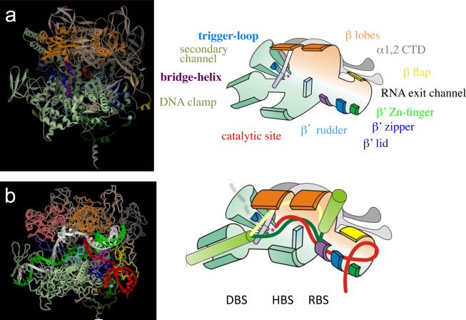
RNA polymerase (RNAP) is a complex molecular machine that governs gene expression and its regulation in all cellular organisms. To accomplish its function of accurately producing a full-length RNA copy of a gene, RNAP performs a plethora of chemical reactions and undergoes multiple conformational changes in response to cellular conditions. At the heart of this machine is the active center, the engine, which is composed of distinct fixed and moving parts that serve as the ultimate acceptor of regulatory signals and as the target of inhibitory drugs. Recent advances in the structural and biochemical characterization of RNAP explain the active center at the atomic level and enable new approaches to understanding the entire transcription mechanism, its exceptional fidelity and control.


Figures

Figure 1
Figure 1. Catalytic activities of RNAP
Schematic representation of enzymatic reactions performed by the EC in different states. i and i+1 represent the product and substrate binding sites of the catalytic center, respectively. Red arrows indicate the direction of the nucleophilic attack. P - phosphate group, N – nucleotide residue, solid lines represent the nascent RNA.
Figure 2
Figure 2. Structural overview of the EC
(a) High-resolution crystal structure of Thermus aquaticus RNAP core (146) (left panel) is shown as colored ribbons (αI, light grey; αII, grey; β, light brown; β', light green; ω, dark green; σ, magenta, as imaged by the Accelris DS-Visualizer program. Structure represents the main channel view of RNAP. Mobile elements are shown as follows: β' BH (residues I705-V835 of T.t. β'), purple; β' TL (T1234-L1256), blue; β' zipper (V26-E47), dark blue; β' Zn-finger (G51-S83), green; β' lid (R525-S538), light blue; β' rudder (N584-S602), turquoise; β lobe 1 (A29-D133, G337-S392) and β lobe 2 (R142-N330), orange; and β flap (G757-S789), yellow. The RNAP β' catalytic loop (N737-Q744) is colored red and the Mg++ ion is shown as a small red sphere. Right panel shows schematic representation of the RNAP structure as a simplified cartoon with the same color code as in the left panel with all the key structural elements indicated. The catalytic center is shown as a small red star. (b) Structural model of the EC (left panel) based on the T.t. EC structure (129). Right panel shows the schematic representation of the structure with the same color code as in the left panel. Duplex DNA is shown as flexible green cylinders; nascent RNA, template and non-template DNA strands are shown as red, dark green and yellow worms, respectively. The main channel secures the RNA-DNA hybrid in the hybrid-binding site (HBS). β' domains clamp the downstream DNA in the DNA duplex binding site (DBS). The β' zipper, Zn-finger and lid along with α flap form the RNA exit channel. RNA-binding site (RBS) is located at the junction of HBS and the RNA exit channel and also functions as rear zip-lock that separates the nascent transcript from the hybrid (see the text for details). The front zip-lock formed by the BH and other elements of the catalytic center separates RNA (shown as a dashed red line) from the hybrid during backtracking, directing it into the secondary channel (shown as a funnel). Transcription goes from right to left.
Figure 3
Figure 3. Structure of the RNAP catalytic center
Left panel shows a close-up view of the catalytic center, which is obtained using high-resolution structures of the T.t. EC in the presence of the non-hydrolizable substrate analog AMPcPP (128). Right panel is a schematics of the left panel indicating specific interactions between evolutionarily conserved amino acid residues, substrate NTP, Mg++ ions and parts of the nucleic acid scaffold, based on the structures of T.t. and yeast ECs (136,55,128,129). Residue numbers corresponding to T.t. β and β' polypeptides are shown. Residues of the Rpb1 of yeast RNAP II that differ from T.t. β' are shown in parenthesis. Three principal groups of residues are indicated: those that coordinate Mg++ ions, those that participate in binding and proper orientation of the NTP α-, β- and γ-phosphates, and those responsible for recognition of correct NTPs. The two Mg++ ions play an essential role in RNA catalysis (114, 110). The first Mg++ (Mg-A) is bound by three aspartates of the β' catalytic loop (D739, D741 and D743). The second Mg2+ (Mg-B) is less stably bound in the enzyme mostly by one aspartate of the β' catalytic loop (D739) via direct interactions, and by one aspartate of β (D686) via water-mediated interactions (128). Solid and dashed lines indicate direct and water-mediated contacts, respectively. During RNA synthesis (or pyrophosphorolysis) Mg-B is involved in the coordination of the NTP α-, β- and γ-phosphates, insuring their optimal spatial alignment for the nucleophilic attack of the activated 3'-OH group on the α-phosphate (shown by the green arrow on right panel). Additionally, the TL residues R1029 and R1239, and also β R557 and R879 bind the NTP β- and γ-phosphates via hydrogen bonding and electrostatic interactions (128). β' H1042 plays an especially important role in the binding of the α- phosphate (128) (and possibly the β-phosphate [136]) and facilitating the nucleophilic attack of the 3'-OH group. To ensure incorporation of correct nucleotides into the RNA, TL M1238 and BH β'T1088 make hydrophobic/van der Waals contacts with the NTP base and the DNA base at the i+1 position, while β' N737 and R704 establish hydrogen bonds with the 2'- and 3'-OH groups of the ribose ring of the NTP (128,50].
Figure 4
Figure 4. Nucleotide addition cycle (NAC) and the dynamics of the catalytic center
(a) Schematic diagram depicting conformational changes in the TL and BH during NTP incorporation and subsequent translocation (14,116). DNA template and RNA strands are shown in blue and red, respectively. The BH is shown in grey and the TL is in blue. Magenta circles indicate catalytic Mg-A (I) and Mg-B (II). In the initial, post-translocated (active) EC, the TL is in the unfolded, “open” conformation (136,128). The binding of the correct NTP (orange) leads to refolding of the TL into an extended α-helical conformation (see panel b), which, in turn stabilizes NTP binding through interactions with the TL. Since NTP binding in the i+1 site stabilizes the EC in the post-translocated state, the substrate serves as a stationary pawl in the ratchet mechanism of RNAP translocation (7). After incorporation of the NTP into RNA and release of pyrophosphate, the TL/BH unit may oscillate between the unfolded (open) conformation and intermediate state in which the TL wedges into the BH (see panel c), forcing the latter to “push” against the RNA-DNA hybrid, thereby inducing translocation of the EC to an active, post-translocated state. According to this model, the TL/BH unit serves as a second, reciprocal pawl, facilitating RNAP translocation in the absence of a substrate (34,7,14). α-amanitin (yellow) and steptolydigin antibiotics have been proposed to interfere with NAC by either interrupting TL-dependent loading of a substrate (50,120) or inhibiting TL/BH-dependent RNAP translocation (14). For further details, refer to the text. (b) TL refolding in the T.t. EC (128). BH (gray cartoon) is packed against the TL refolded as trigger-helices (blue cartoon) as a result of NTP binding (i+1, insertion site, orange sticks). i site RNA is shown as red sticks. Trigger-helixes residues interacting with NTP (Met1238, Arg1239, Phe1241, and His 1242) are shown as blue sticks. (c) Distortion of the yeast RNAPII BH (grey cartoon) by the wedging TL (cartoon blue) in the presence of α-amanitin (sticks yellow) (14). i site RNA is shown as red sticks, wedging residue of the TL, Leu1081 is shown as teal sticks, interacting with α-amanitin His1085 is shown as magenta sticks (14,50).
Figure 5
Figure 5. Structural model of the trapped intermediate during termination
RNAP structure with the explanatory scheme represents the last stage of the termination process (40,31). Key structural elements are color coded (see Figure 2b): gray (green) – non-template (template) strand of DNA; red – nascent RNA; yellow - β flap; violet – β' lid; blue – β' zipper; green – Zn-finger; rose and gold - β lobes 1 and 2; aqua - β rudder; dark blue - TL; teal – DNA clamp and other parts of β'. Star - the catalytic center. The termination RNA hairpin stem grows to its final size of 7–8 bp invading the primary channel, unwinding the upstream portion of the hybrid and initiating collapse of the upstream edge of the transcription bubble. The head of the hairpin bends around the downstream edge of the transcription bubble, clashing with the TL. This may force the TL to move towards the active site and permanently fold into a “closed” conformation. Additionally, the hairpin action results in disruption of interactions in the RBS and HBS and distortion of the hybrid, which could cause complete and irreversible enzyme inactivation. At the same time, the TL movement induces DNA-binding-clamp (DBS) opening, resulting in a simultaneous release of both the nascent RNA and the DNA template.

References

    1. Abbondanzieri E, et al. Direct observation of base-pair stepping by RNA polymerase. Nature. 2005;438:460–465. - PMC - PubMed
    1. Adelman K, et al. Molecular mechanism of transcription inhibition by peptide antibiotic Microcin J25. Mol. Cell. 2004;14:753–762. - PubMed
    1. Altmann CR, Solow-Cordero DE, Chamberlin MJ. RNA cleavage and chain elongation by Escherichia coli DNA-dependent RNA polymerase in a binary enzyme. RNA complex. Proc Natl Acad Sci U S A. 1994;91:3784–8. - PMC - PubMed
    1. Archambault J, Friesen J. Genetics of RNA polymerases I, II and III. Microbiol Rev. 1993;57:703–724. - PMC - PubMed
    1. Artsimovitch I, et al. Allosteric modulation of the RNA polymerase catalytic reaction is an essential component of transcription control by rifamycins. Cell. 2005;122:351–63. - PubMed

Publication types

MeSH terms

Substances

LinkOut - more resources