Review
. 2009:78:363-97.
doi: 10.1146/annurev.biochem.78.082307.091526.

Regulation and cellular roles of ubiquitin-specific deubiquitinating enzymes

Affiliations
Review

Regulation and cellular roles of ubiquitin-specific deubiquitinating enzymes

Francisca E Reyes-Turcu et al. Annu Rev Biochem. 2009.

Abstract

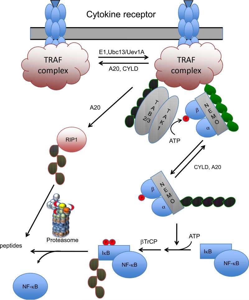
Deubiquitinating enzymes (DUBs) are proteases that process ubiquitin or ubiquitin-like gene products, reverse the modification of proteins by a single ubiquitin(-like) protein, and remodel polyubiquitin(-like) chains on target proteins. The human genome encodes nearly 100 DUBs with specificity for ubiquitin in five gene families. Most DUB activity is cryptic, and conformational rearrangements often occur during the binding of ubiquitin and/or scaffold proteins. DUBs with specificity for ubiquitin contain insertions and extensions modulating DUB substrate specificity, protein-protein interactions, and cellular localization. Binding partners and multiprotein complexes with which DUBs associate modulate DUB activity and substrate specificity. Quantitative studies of activity and protein-protein interactions, together with genetic studies and the advent of RNAi, have led to new insights into the function of yeast and human DUBs. This review discusses ubiquitin-specific DUBs, some of the generalizations emerging from recent studies of the regulation of DUB activity, and their roles in various cellular processes.


Figures

Figure 1
Figure 1
Domain structure of yeast UBP deubiquitinating enzymes. The catalytic core is indicated by the blue boxes. Other common domains are indicated with differently colored boxes: MATH, meparin and TRAF homology; Rhodanese, rhodanese-like; ZnF-UBP, zinc finger common in UBP DUBs, UBL, ubiquitin-like, and UBA, ubiquitin-associated. Three sites of common insertions within the catalytic core are indicated by arrows. These insertions and the N- and C-terminal extensions are thought to provide specific sequences that define substrate specificity and provide interaction surfaces for binding to adapters and scaffolds.
Figure 2
Figure 2
Substrate induced conformational changes upon ubiquitin binding to DUBs. A) a loop occluding the active must be displaced to allow substrate binding and access to the active site. B) A larger domain occluding the active site must be displaced upon substrate binding or association with a scaffold protein. C) The binding of the substrate induces a conformational change in the catalytic triad allowing it to achieve a catalytically competent geometry.
Figure 3
Figure 3
Proteasome bound deubiquitinating enzymes. Deubiquitinating enzymes are indicated by red crescents, the substrate as a green line, and ubiquitin as blue ovals. POH1 catalyzes the release of a polyubiquitin chain “en bloc” as the substrate is engaged and translocated through the gated pore of the 20S protease. RPN10 (yellow) binds the polyubiquitin chain and the distal end of the chain can be removed by the action of UCH37 bound to ADRM1 (orange). USP14 is bound to the proteasome via interactions with RPN1 (purple) and probably removes mono ubiquitin attached to the substrate.
Figure 4
Figure 4
Deubiquitination of histones H2A and H2B regulate chromatin structure and activity. Panel A details the dynamic ubiquitination/deubiquitination of H2B in yeast. These DUBs are localized to sites of action by interaction with other chromatin bound proteins that act as scaffolds and activate the DUB activity. Panel B shows the ubiquitination/deubiquitination of H2A in higher eukaryotes. A putative binding partner for UBP16 with a role in chromatin condensation is shown with question marks.
Figure 5
Figure 5
A generalized model for signaling through the NF-κB pathway. Upon ligand binding to receptors for Tumor Necrosis Factor, Interleukins, Toll ligand, or T- and B-cell antigens a signaling complex is assembled. This complex contains TRAFs (TNF receptor associated factors) that catalyze the formation of a K63-linked polyubiquitin chain (green) on themselves and/or other proteins in the complex. The K63-linked polyubiquitin then recruits the TAB2/3-TAK1 kinase and its substrate IκB kinase (consisting of kinases IKKα, IKKβ, and the regulatory subunit NEMO). Subsequently the phosphorylation of IKKβ and the K63 polyubiquitination of NEMO activates the IKK activity. IKK phosphorylates IκB triggering its K48 polyubiquitination (red) by βTRCP and its degradation by the proteasome. The liberated NF-κB then enters the nucleus and acts as a transcription factor activating genes involved in inflammation and immune responses. The signaling is downregulated by two K63 specific DUBs, A20 and CYLD and defects in either lead to prolonged signaling. RIP1, a component of the TRAF complex assembled upon TNFR1 signaling is one of the targets for K63 polyubiquitination. K63 polyubiquitinated RIP1 is deubiquitinated by A20. This DUB is also a ubiquitin ligase that then assembles a K48 polyubiquitin chain on RIP1 leading to its degradation by the proteasome.

References

    1. Hershko A, Ciechanover A. The ubiquitin system. Annu Rev Biochem. 1998;67:425–79. - PubMed
    1. Hicke L. Protein regulation by monoubiquitin. Nat Rev Mol Cell Biol. 2001;2:195–201. - PubMed
    1. Pickart CM, Eddins MJ. Ubiquitin: structures, functions, mechanisms. Biochim Biophys Acta. 2004;1695:55–72. - PubMed
    1. Pickart CM, Fushman D. Polyubiquitin chains: polymeric protein signals. Curr Opin Chem Biol. 2004;8:610–6. - PubMed
    1. Pickart CM. Mechanisms underlying ubiquitination. Annu Rev Biochem. 2001;70:503–33. - PubMed

LinkOut - more resources