Review
. 2009:78:515-40.
doi: 10.1146/annurev.biochem.77.060806.091251.

Lipid-dependent membrane protein topogenesis

Affiliations
Review

Lipid-dependent membrane protein topogenesis

William Dowhan et al. Annu Rev Biochem. 2009.

Abstract

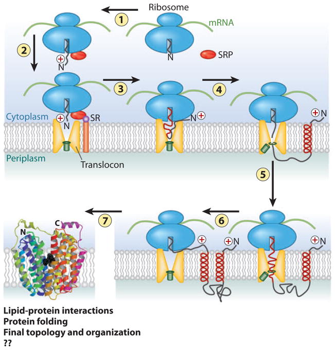
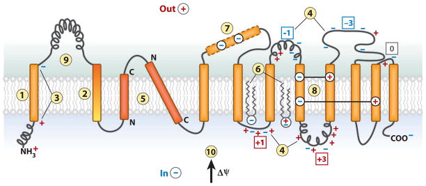
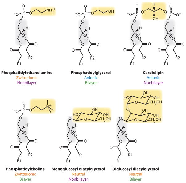
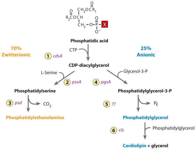
The topology of polytopic membrane proteins is determined by topogenic sequences in the protein, protein-translocon interactions, and interactions during folding within the protein and between the protein and the lipid environment. Orientation of transmembrane domains is dependent on membrane phospholipid composition during initial assembly as well as on changes in lipid composition postassembly. The membrane translocation potential of negative amino acids working in opposition to the positive-inside rule is largely dampened by the normal presence of phosphatidylethanolamine, thus explaining the dominance of positive residues as retention signals. Phosphatidylethanolamine provides the appropriate charge density that permits the membrane surface to maintain a charge balance between membrane translocation and retention signals and also allows the presence of negative residues in the cytoplasmic face of proteins for other purposes.


Figures

Figure 1
Figure 1
Overview of membrane protein synthesis, membrane insertion, and assembly. 1. As the N-terminal domain of a nascent polypeptide chain emerges from the ribosome, it complexes with the signal recognition particle (SRP). 2. The resulting complex is delivered to the membrane in association with the translocon through binding with the SRP receptor (SR). 3. Translation proceeds with threading of the nascent chain into the translocon (SecYEG in prokaryotes and Sec61 in eukaryotes) pore, which is sealed by the plug (green rectangle). YidC (not shown) in bacteria plays a dual role with or independent of the translocon in the integration and assembly of a subset inner membrane proteins. 4. Translation proceeds with the synthesis of the first transmembrane domain (TM), which exits laterally as an α-helix into the lipid bilayer with retention of the positively charged N-terminal domain (according to the positive-inside rule) on the cytoplasmic side of the membrane. 5. The plug opens to allow the next extramembrane domain to cross the membrane and exit into the periplasm. 6. The next TM enters the translocon channel followed by lateral exit into the membrane and cytoplasmic orientation of the next extramembrane domain. Steps 4 through 6 are repeated until synthesis is complete. 7. As TMs enter the membrane, lipid-protein interactions, short-range protein-protein interactions, and long-range helix packing now govern topological and folding events [in a coordinated but undefined (??) manner], resulting in a final compact native structure. In eukaryotes, the periplasm corresponds to the lumen of the endoplasmic reticulum. Figure was adapted and extensively modified from Reference . Folded protein is lactose permease (LacY) of Escherichia coli from Reference and reprinted with permission from American Association for the Advancement of Science.
Figure 2
Figure 2
The features of polytopic membrane proteins and biological membranes that determine folding and final transmembrane domain (TM) topology. TM integration is driven by (1) the hydrophobicity of TMs, and initial orientation is influenced by (2) the hydrophobic gradient (highest outward) along a TM, (3) the charge difference or (4) the positive-inside rule, (5) preferential orientation of highly hydrophobic TMs with the C terminus out if short and the N terminus out if long. After exit from the translocon, (6) final topology is influenced by lipid headgroup-protein charge interactions. During protein folding, interactions between TMs can stabilize (7) a domain containing charged residues as (8) a TM by compensating salt bridges. (9) A rapidly folding extramembrane domain can prevent reorientation of TMs during late folding events. (10) The positive outward membrane potential favors translocation of net negative domains and retention of net positive domains.
Figure 3
Figure 3
Structure and physical chemical properties of lipids. Stick diagram of the carbon backbone of lipids with R substituting for the long hydrocarbon portion of fatty acids. Phospholipids found normally in E. coli are in the top row. Foreign lipids that have been introduced into E. coli mutants lacking phosphatidylethanolamine are in the bottom row. The charge nature of the lipid headgroups is noted. Depending on fatty acid composition, solvent, and temperature, those lipids that assume nonbilayer organization in solution are indicated in contrast to those lipids that only form bilayer structures.
Figure 4
Figure 4
Pathway for synthesis of the major phospholipids of E. coli. The following enzymes with their respective genes indicated are 1. CDP-diacylglycerol synthase; 2. phosphatidylserine synthase; 3. phosphatidylserine decarboxylase; 4. phosphatidylglycerol-3-P synthase; 5. phosphatidylglycerol-3-P phosphatase; 6. cardiolipin synthase. The X in phosphatidic acid is an OH and is in the position that changes depending on the downstream pathways. The remaining 5% of total phospholipids are primarily the minor lipids (black).
Figure 5
Figure 5
Topological organization of LacY as a function of membrane lipid composition. (a) The topology of LacY initially assembled in E. coli with wild-type phospholipid composition is shown. Rectangles define the transmembrane domains (TMs) (79), oriented with the cytoplasm above the figure. TMs (Roman numerals), extramembrane domains [P for periplasmic and C for cytoplasmic as oriented in phosphatidylethanolamine (PE)-containing cells], N terminus (NT), and C terminus (CT) are indicated. The net charge of each extramembrane domain is indicated next to the domain name. The approximate locations of negatively charged (blue) and positively charged (red ) residues are indicated. (b) A funnel-shaped energy landscape that defines the folding pathway of a membrane protein such as LacY in wild-type cells is depicted. The horizontal axis ( funnel circumference) represents the conformational space occupied by protein as it folds, and the vertical axis represents the internal free energy of a given polypeptide conformation. As TMs I–XII fold, they move down the funnel to the lowest energy state as defined by the final organization of native LacY. (c) The topology of LacY assembled in E. coli cells lacking PE is shown. TMs I–VI are inverted with respect to TMs VIII–XII, which still exhibit native topology. TM VII (red ) is exposed to the periplasm, resulting in breakage of the salt bridges with TM X and TM XI. (d ) A new energy landscape defines the energetics of folding in PE-lacking cells of the two halves of LacY with the N-terminal bundle forming a new energy minimum separated by a large activation energy, which disconnects it from the native state. (e) The topological organization of LacY after synthesis of PE in cells after assembly of LacY. All of LacY assumes a native topological organization except the domain defined by NT-TM I-P1-TM II. ( f ) The presence of PE raises the energy minimum (arrow) of the inverted topology of TM III–VII, resulting in low activation energy for a return to the native topology. A large energy barrier remains for TM I flipping with a new miniloop structure (TM II) allowing all domains to exist at their respective energy minima.
Figure 6
Figure 6
Phosphatidylethanolamine (PE) and the positive-inside rule. (left) A cytoplasmic domain is shown containing a mixture of negative and positive amino acids. PE is shown to suppress or neutralize the presence of negative residues ( yellow), which increases the effective positive charge potential, thus favoring retention on the cytoplasmic side of the membrane. (right) In the absence of PE, negative residues exert their full potential and result in translocation of a domain that exhibits a lower effective net positive charge. The proton motive force (arrow) positive outward determines domain directional movement depending on the domain effective charge. Postassembly addition of PE changes the effective net charge of these domains and favors reorientation.
Figure 7
Figure 7
Distribution of charged amino acids in homologous sugar permeases. Sequence alignments are shown for E. coli (Ec) lactose (LacY), raffinose (RafB), and sucrose (CscB) permeases and for Klebsiella pneumoniae (Kp) and Citrobacter freundii (Cf ) lactose permeases. Positively or negatively charged amino acids are colored red or blue, respectively.
Figure 8
Figure 8
Distribution of charged amino acids in homologous amino acid permeases. Sequence alignments shown for E. coli γ-aminobutyrate permease (GabP), phenylalanine (PheP), lysine (LysP), and aromatic (AroP) permeases. Positively or negatively charged amino acids are colored with red or blue, respectively, and aromatic residues in TM III are colored purple.

References

    1. Driessen AJ, Nouwen N. Protein translocation across the bacterial cytoplasmic membrane. Annu Rev Biochem. 2008;77:643–67. - PubMed
    1. Rapoport TA. Protein translocation across the eukaryotic endoplasmic reticulum and bacterial plasma membranes. Nature. 2007;450:663–69. - PubMed
    1. Pitonzo D, Skach WR. Molecular mechanisms of aquaporin biogenesis by the endoplasmic reticulum Sec61 translocon. Biochim Biophys Acta. 2006;1758:976–88. - PubMed
    1. von Heijne G. Membrane-protein topology. Nat Rev Mol Cell Biol. 2006;7:909–18. - PubMed
    1. Sadlish H, Pitonzo D, Johnson AE, Skach WR. Sequential triage of transmembrane segments by Sec61α during biogenesis of a native multispanning membrane protein. Nat Struct Mol Biol. 2005;12:870–88. - PubMed

Publication types

MeSH terms

LinkOut - more resources