. 2009 Jul;276(13):3602-17.
doi: 10.1111/j.1742-4658.2009.07080.x. Epub 2009 May 20.

Analysis of Lsm1p and Lsm8p domains in the cellular localization of Lsm complexes in budding yeast

Affiliations
Free PMC article

Analysis of Lsm1p and Lsm8p domains in the cellular localization of Lsm complexes in budding yeast

Martin A M Reijns et al. FEBS J. 2009 Jul.
Free PMC article

Abstract

In eukaryotes, two heteroheptameric Sm-like (Lsm) complexes that differ by a single subunit localize to different cellular compartments and have distinct functions in RNA metabolism. The cytoplasmic Lsm1-7p complex promotes mRNA decapping and localizes to processing bodies, whereas the Lsm2-8p complex takes part in a variety of nuclear RNA processing events. The structural features that determine their different functions and localizations are not known. Here, we analyse a range of mutant and hybrid Lsm1 and Lsm8 proteins, shedding light on the relative importance of their various domains in determining their localization and ability to support growth. Although no single domain is either essential or sufficient for cellular localization, the Lsm1p N-terminus may act as part of a nuclear exclusion signal for Lsm1-7p, and the shorter Lsm8p N-terminus contributes to nuclear accumulation of Lsm2-8p. The C-terminal regions seem to play a secondary role in determining localization, with little or no contribution coming from the central Sm domains. The essential Lsm8 protein is remarkably resistant to mutation in terms of supporting viability, whereas Lsm1p appears more sensitive. These findings contribute to our understanding of how two very similar protein complexes can have different properties.


Figures
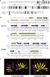
Fig. 1
Fig. 1
Structural features of Lsm1 and Lsm8 polypeptides. (A) Alignment of Lsm1p and Lsm8p using clustal w [48] (B) 2D structure predictions for Lsm1p and Lsm8p using 3D-PSSM [32]. Arrowheads indicate sites of N- and C-terminal deletions and fusions. * indicates residues forming putative RNA-binding Knuckle motifs. Green boxes indicate regions that are predicted to form α helices (H), blue arrows indicate regions that are predicted to form β strands (E) and lines indicate regions that are predicted to form random coil (C). Numbers indicate the confidence scores of these predictions for each residue, with 5–9 (in bold) indicating high confidence. (C) 3D structural prediction for Lsm1p and Lsm8p, made using default settings of swiss-model [49]. The model shown for Lsm1p covers residues 44–155 and is based on homology to a Sm-like archaeal protein from Pyrobaculum aerophilum (1m5q) [40]. The model shown for Lsm8p covers residues 1–67 and is based on homology to a heptameric Sm protein from P. aerophilum (1i8f) [50]. Arrows indicate break-points for our hybrids; the green arrow for Lsm8p indicates residue 67, whereas the break-point for our hybrids is residue 73; putative RNA-binding residues are shown in red.
Fig. 2
Fig. 2
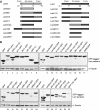
Lsm1p and Lsm8p mutant and hybrid proteins are stably produced. (A) Schematic overview of hybrids and deletion mutants of Lsm1p and Lsm8p. (B) MPS26 cells with plasmids expressing GFP-tagged hybrid and mutant proteins (Table S1) were grown in SD–Ura–Met (or SD–Ura+Met; lane 31) and aliquots of total protein from equal D600 units of cells were separated by SDS/PAGE and western blotted, probing with anti-GFP IgG2a. Hybridization with anti-(α-tubulin) IgG1 assesses equivalence of loading. Lsm8 rna mutants carry point mutations in putative RNA-binding residues (for details of all mutants and hybrids see Table S1). Additional bands in lanes 27 and 29 likely represent cleaved off GFP.
Fig. 3
Fig. 3
Lsm1p and Lsm8p N- and C-terminal extensions do not suffice for localization of GFP to P-bodies or nuclei. Strain MPS26 was transformed with pGFP–N-LSM1 (Lsm1), pMR144 (1N), pMR133 (1C), pGFP–N-LSM8 (Lsm8), pMR132 (8N), pMR156 (8C) or pGFP–N-FUS (GFP; Table S1). Cells were grown in SD–Ura–Met and localization was examined during log phase growth or 10 min after hypo-osmotic shock (for Lsm1p only). The images shown in this and all other figures are representative of the majority of cells in each given experiment.
Fig. 4
Fig. 4
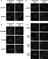
Effects of mutations in Lsm8p on its nuclear localization. (A) Lsm8p C-terminal domain mutations. (B) Lsm8p N-terminal domain mutations and recombinant Lsm1p containing the Lsm8p N-terminal 10 amino acids. (C) Sm domain replacements; see Fig 2A for an explanation of the constructs. (D) Mutations in or near the putative RNA-binding residues of Knuckle I and II. MPS26 was transformed with plasmids (A) pMR70, pMR80, pMR84 and pMR104; (B) pMR117, pMR126, pMR140, pMR141, pMR115 and pMR124; (C) pMR114, pMR116, pMR123 and pMR125; and (D) pMPS8, pMR76, pMR77, pMR78, pMR83, pMR92, pMR93 and pMR94 (see Table S1 for plasmid descriptions). Cells were grown in SD–Ura(–Met) and localization was examined in live cells during log-phase growth. We show the results for live cells only because we found that nuclear localization of our GFP-tagged proteins, including that of GFP–Lsm8, was significantly reduced after fixing (either using 4% formaldehyde or methanol). Intensities of nuclear and cytoplasmic signals were measured using imagej 1.38w and the average ratios of nuclear/cytoplasmic signals are indicated within each image. Where no nuclear accumulation was detected, a ratio of 1.0 is given.
Fig. 5
Fig. 5
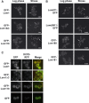
No single Lsm1p domain is essential for localization to P-bodies. (A) Lsm1p C-terminal domain mutations. (B) Lsm1p N-terminal domain mutations or central Sm domain replacement. See Fig 2A for an explanation of the constructs. Arrows indicate P-bodies; * indicate nuclear accumulation. (C) Lsm1p, Lsm1ΔCp and Lsm118p colocalize with Dcp2p in foci. AEMY25 (lsm1Δ) was transformed with plasmids: (A) pGFP–N-LSM1, pMR69 and pMR79; (B) pMR124, pMR126, pMR135 and pMR123; (C) pGFP–N-LSM1, pMR69, pMR79 or pGFP–N-FUS together with pRP1155 (DCP2–RFP; see Table S1 for plasmid descriptions). Cells were grown in SD–Ura(–Met) (A,B) or SD–Ura–Leu–Met (C) and localization was examined in live cells during log phase growth, after hypo-osmotic shock (stress; A,B) or after glucose starvation (C). Approximate percentages of cells showing focal accumulation of GFP signals after stress are given with each of the images in (A) and (B).
Fig. 6
Fig. 6
The Lsm1p and Lsm8p N-terminal domains are required for distinct localization. MPS26 was transformed with plasmids: (A) pMR129, pMR130, pMR137 and pMR138; (B) pMR143, pMR145, pMR147 and pMR148; (C) pMR134, pMR135, pMR140 and pMR141; (D) pMR150, pMR151, pMR153 and pMR154 (see Fig 2A for an explanation of the constructs and Table S1 for plasmid descriptions). Cells were grown in SD–Ura–Met to D600 = 1–2, and localization was examined in live cells. Nuclei are indicated by *, cytoplasmic foci are indicated by arrows. Intensities of nuclear and cytoplasmic signals were measured using imagej 1.38w and the average ratios of nuclear/cytoplasmic signals are indicated within each image. Where no nuclear accumulation was detected, a ratio of 1.0 is given.
Fig. 7
Fig. 7
Correlation between viability and correct localization of Lsm1 and Lsm8 hybrid and mutant proteins. (A) Mutant proteins that accumulate in the nucleus. (B) Mutant proteins that accumulate in P-bodies. Viability was scored by comparison with the wild-type plasmid (++++) and the GFP only negative control (−). Proteins that accumulate both in nuclei and P-bodies are indicated by *. For a more detailed scoring of growth phenotypes for all different constructs see Tables S2 and S3.
Fig. 8
Fig. 8
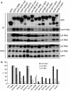
Analysis of complex formation and U6 snRNA binding of Lsm8 mutant and hybrid proteins. MPS26 cells carrying the appropriate plasmids were grown in SD–Ura–Leu–Met at 23 °C. Proteins were immunoprecipitated with affinity-purified rabbit anti-GFP. (A) Recombinant GFP-tagged protein and genomically encoded, co-precipitated Lsm7–Myc were visualized by western blotting; coprecipitated U4 and U6 snRNA, and total U6, U4 snRNA and scR1 present in the extracts were analysed by northern blotting. (B) Coprecipitated levels of Lsm7–Myc protein, U6 and U4 snRNA were quantified using imagequant software (Molecular Dynamics), normalized to GFP only background, and plotted as a percentage of wild-type. Immunoprecipitations were performed on two biological replicates, which showed similar results.
Fig. 9
Fig. 9
Levels of U4, U6 and U4/U6 snRNAs in lsm8 mutant and hybrid strains. MPS11, which depends on a CENHIS3 plasmid expressing HA–Lsm8p from the GAL1-10 promoter, was transformed with plasmids expressing the mutant and hybrid proteins from the MET25 promoter. Cells were grown in SDGal–Ura at 30 °C to mid-log phase before shifting them to SD–Ura–Met and growing for an additional 10 h at 30 °C. After 10 h equal numbers of cells were harvested for each of the mutants and total RNA was extracted under nondenaturing conditions. (A) Northern probed for U6 RNA after nondenaturing PAGE of total RNA. U1 snRNA was used as a loading control. (B) The same northern probed for U4 RNA. (C) Western blot on total protein probed with α-HA antibody confirms that cells are almost entirely depleted of HA–Lsm8p after 10 h of growth on glucose; similar levels of α-tubulin confirm equal loading. Quantifications on northern images were performed using imagequant; relative levels of U4, U6 and U4/U6 were corrected for U1 loading and expressed as a percentage of the level in the GFP–LSM8 positive control. Total U4 (U4 tot.) amounts, i.e. U4 present on its own as well as annealed to U6, were quantified in (B).

Similar articles

Cited by

References

    1. Mayes AE, Verdone L, Legrain P, Beggs JD. Characterization of Sm-like proteins in yeast and their association with U6 snRNA. EMBO J. 1999;18:4321–4331. - PMC - PubMed
    1. Salgado-Garrido J, Bragado-Nilsson E, Kandels-Lewis S, Séraphin B. Sm and Sm-like proteins assemble in two related complexes of deep evolutionary origin. EMBO J. 1999;18:3451–3462. - PMC - PubMed
    1. Achsel T, Brahms H, Kastner B, Bachi A, Wilm M, Lührmann R. A doughnut-shaped heteromer of human Sm-like proteins binds to the 3′-end of U6 snRNA, thereby facilitating U4/U6 duplex formation in vitro. EMBO J. 1999;18:5789–5802. - PMC - PubMed
    1. Pannone BK, Xue D, Wolin SL. A role for the yeast La protein in U6 snRNP assembly: evidence that the La protein is a molecular chaperone for RNA polymerase III transcripts. EMBO J. 1998;17:7442–7453. - PMC - PubMed
    1. Spiller MP, Boon KL, Reijns MAM, Beggs JD. The Lsm2-8 complex determines nuclear localization of the spliceosomal U6 snRNA. Nucleic Acids Res. 2007;35:923–929. - PMC - PubMed

Publication types

MeSH terms