Lines of action and stabilizing potential of the shoulder musculature
- PMID: 19490400
- PMCID: PMC2740966
- DOI: 10.1111/j.1469-7580.2009.01090.x
Lines of action and stabilizing potential of the shoulder musculature
Abstract
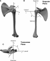
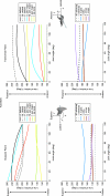
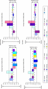
The objective of the present study was to measure the lines of action of 18 major muscles and muscle sub-regions crossing the glenohumeral joint of the human shoulder, and to compute the potential contribution of these muscles to joint shear and compression during scapular-plane abduction and sagittal-plane flexion. The stabilizing potential of a muscle was found by assessing its contribution to superior/inferior and anterior/posterior joint shear in the scapular and transverse planes, respectively. A muscle with stabilizing potential was oriented to apply more compression than shear at the glenohumeral joint, whereas a muscle with destabilizing potential was oriented to apply more shear. Significant differences in lines of action and stabilizing capacities were measured across sub-regions of the deltoid and rotator cuff in both planes of elevation (P < 0.05), and substantial differences were observed in the pectoralis major and latissimus dorsi. The results showed that, during abduction and flexion, the rotator cuff muscle sub-regions were more favourably aligned to stabilize the glenohumeral joint in the transverse plane than in the scapular plane and that, overall, the anterior supraspinatus was most favourably oriented to apply glenohumeral joint compression. The superior pectoralis major and inferior latissimus dorsi were the chief potential scapular-plane destabilizers, demonstrating the most significant capacity to impart superior and inferior shear to the glenohumeral joint, respectively. The middle and anterior deltoid were also significant potential contributors to superior shear, opposing the combined destabilizing inferior shear potential of the latissimus dorsi and inferior subscapularis. As potential stabilizers, the posterior deltoid and subscapularis had posteriorly-directed muscle lines of action, whereas the teres minor and infraspinatus had anteriorly-directed lines of action. Knowledge of the lines of action and stabilizing potential of individual sub-regions of the shoulder musculature may assist clinicians in identifying muscle-related joint instabilities, assist surgeons in planning tendon reconstructive surgery, aid in the development of rehabilitation procedures designed to improve joint stability, and facilitate development and validation of biomechanical computer models of the shoulder complex.
Figures


References
-
- Basmajian J, de Luca C. Muscles Alive: Their Functions Revealed by Electromyography. Baltimore: Williams and Wilkins; 1984.
-
- Bigliani LU, Kelkar R, Flatow EL, Pollock RG, Mow VC. Glenohumeral stability. Biomechanical properties of passive and active stabilizers. Clin Orthop. 1996;330:13–30. - PubMed
-
- Charlton IW, Johnson GR. Application of spherical and cylindrical wrapping algorithms in a musculoskeletal model of the upper limb. J Biomech. 2001;34:1209–1216. - PubMed
-
- Chiari L, Della Croce U, Leardini A, Cappozzo A. Human movement analysis using stereophotogrammetry. Part 2: instrumental errors. Gait Posture. 2005;21:197–211. - PubMed
Publication types
MeSH terms
LinkOut - more resources
Full Text Sources
Research Materials

