

Nerve growth factor and receptor expression in rheumatoid arthritis and spondyloarthritis
- PMID: 19490633
- PMCID: PMC2714133
- DOI: 10.1186/ar2716
Nerve growth factor and receptor expression in rheumatoid arthritis and spondyloarthritis
Abstract
Introduction: We previously described the presence of nerve growth factor receptors in the inflamed synovial compartment. Here we investigated the presence of the corresponding nerve growth factors, with special focus on nerve growth factor (NGF).
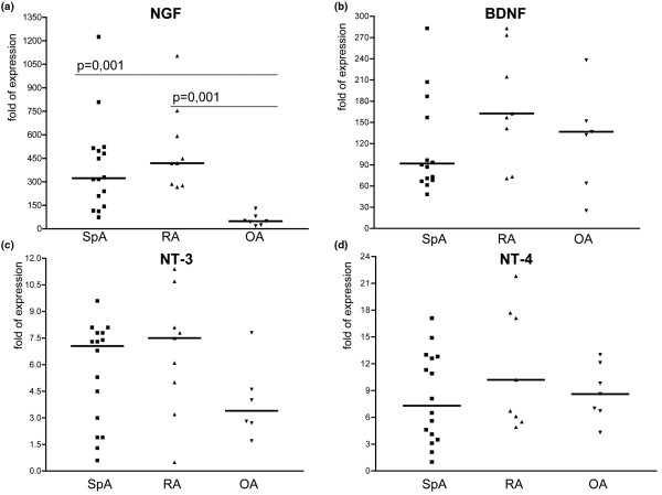
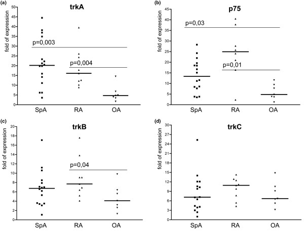
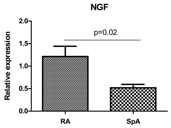
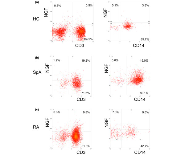
Methods: mRNA expression levels of four ligands (NGF, brain derived growth factor (BDNF), neurotrophin (NT)-3, NT-4) and their four corresponding receptors (tyrosine kinase (trk) A, trkB, trkC, NGFRp75) were determined in the synovial fluid (SF) cells of 9 patients with rheumatoid arthritis (RA) and 16 with spondyloarthritis (SpA) and compared with 7 osteoarthritis (OA) patients. NGF was also determined in synovial tissue (ST) biopsies of 10 RA and 10 SpA patients. The production of NGF by monocytes and lymphocytes was assessed by flow cytometry of SF cells, synovial tissue derived fibroblast-like synoviocytes (FLS) were assessed by ELISA on culture supernatant.
Results: SF cell analysis revealed a clear BDNF and NGF mRNA expression, with significantly higher NGF expression in RA and SpA patients than in the OA group. NGF expression was higher in ST samples of RA as compared to SpA. Using intracellular FACS analysis, we could demonstrate the presence of the NGF protein in the two inflammatory arthritis groups on both CD3+ T lymphocytes and CD14+ cells, i.e. monocytes/macrophages, whereas cultured FLS did not produce NGF in vitro.
Conclusions: Neurotrophins and especially NGF are expressed in the synovial fluid and tissue of patients with peripheral synovitis. The presence of neurotrophins as well as their receptors, in particular the NGF/trkA-p75 axis in peripheral synovitis warrants further functional investigation of their active involvement in chronic inflammatory arthritis.
Figures
References
-
- Tessarollo L. Pleiotropic functions of neurotrophins in development. Cytokine Growth Factor Rev. 1998;9:125–137. - PubMed
-
- Aloe L, Tuveri MA, Carcassi U, Levi-Montalcini R. Nerve growth factor in the synovial fluid of patients with chronic arthritis. Arthritis Rheum. 1992;35:351–355. - PubMed
-
- Pozza M, Guerra M, Manzini E, Calza L. A histochemical study of the rheumatoid synovium: focus on nitric oxide, nerve growth factor high affinity receptor, and innervation. J Rheumatol. 2000;27:1121–1127. - PubMed
-
- Wu Z, Nagata K, Iijima T. Immunohistochemical study of NGF and its receptors in the synovial membrane of the ankle joint of adjuvant-induced arthritic rats. Histochem Cell Biol. 2000;114:453–459. - PubMed
-
- Iannone F, De Bari C, Dell'Accio F, Covelli M, Patella V, Lo Bianco G, Lapadula G. Increased expression of nerve growth factor (NGF) and high affinity NGF receptor (p140 TrkA) in human osteoarthritic chondrocytes. Rheumatology (Oxford) 2002;41:1413–1418. - PubMed
Publication types
MeSH terms
Substances
LinkOut - more resources
Full Text Sources
Other Literature Sources
Medical
Research Materials
