. 2009 Aug;159(2):178-84.
doi: 10.1016/j.jviromet.2009.03.017. Epub 2009 Mar 26.

In vivo biotinylation and capture of HIV-1 matrix and integrase proteins

Affiliations

In vivo biotinylation and capture of HIV-1 matrix and integrase proteins

Michael Belshan et al. J Virol Methods. 2009 Aug.

Abstract

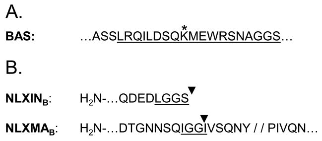
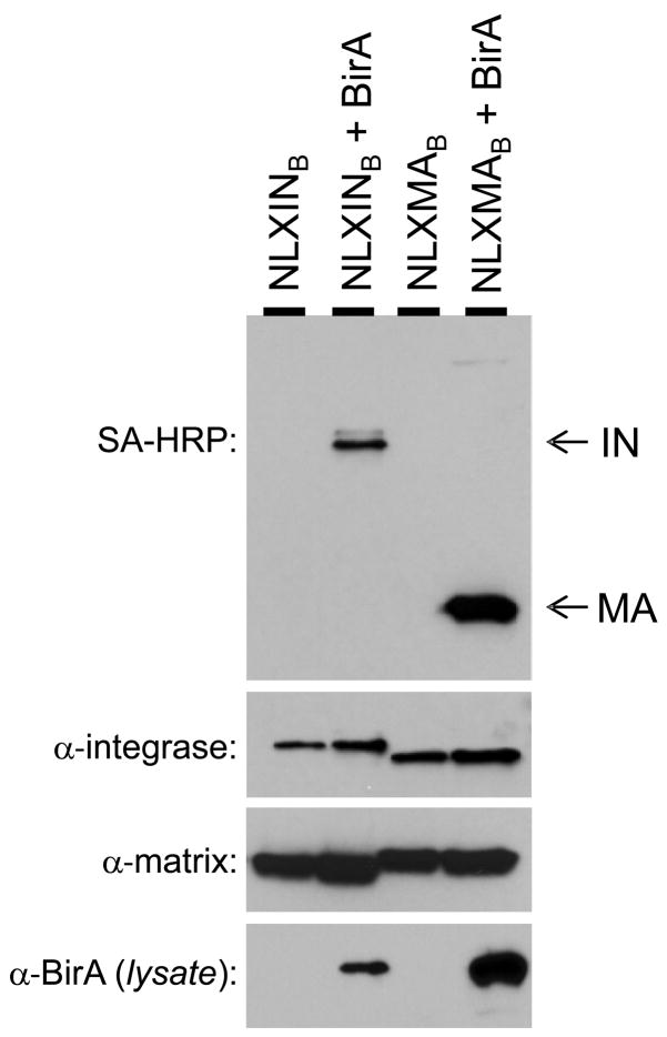
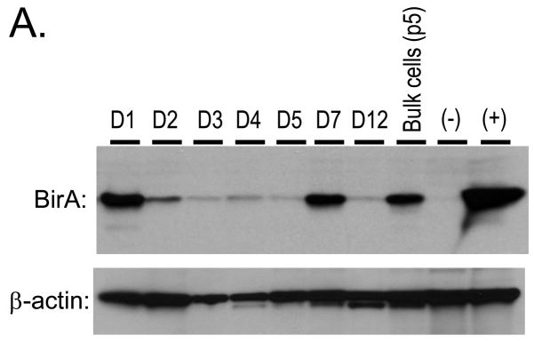
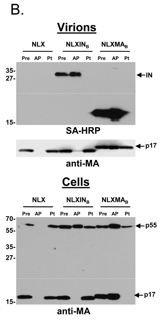
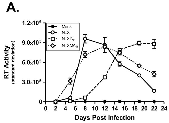
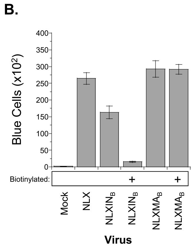
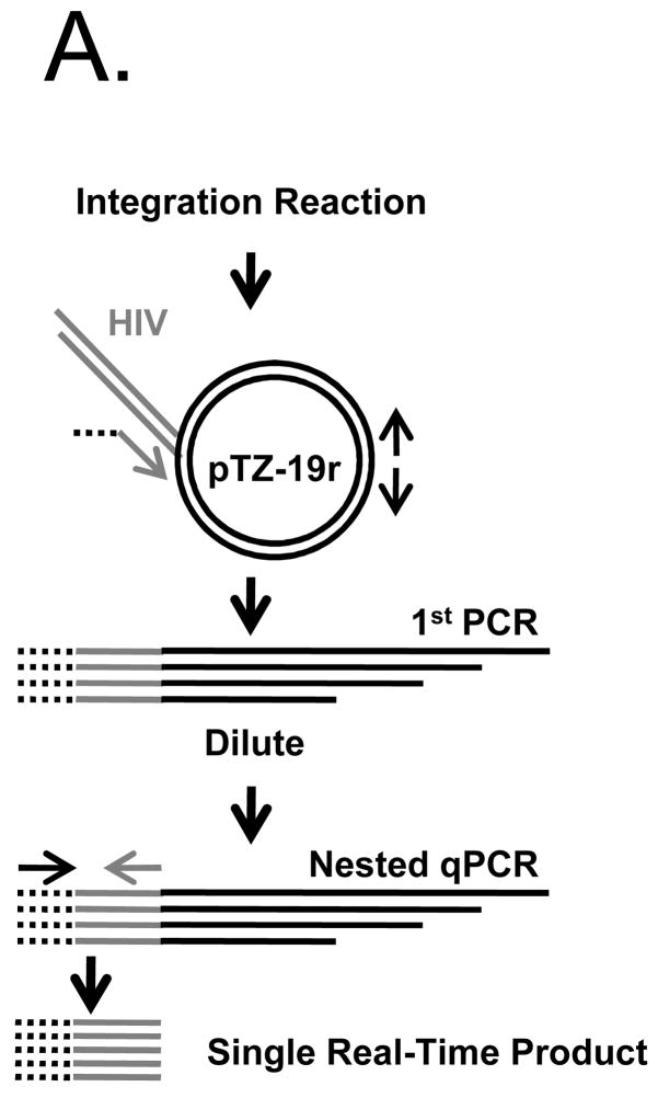
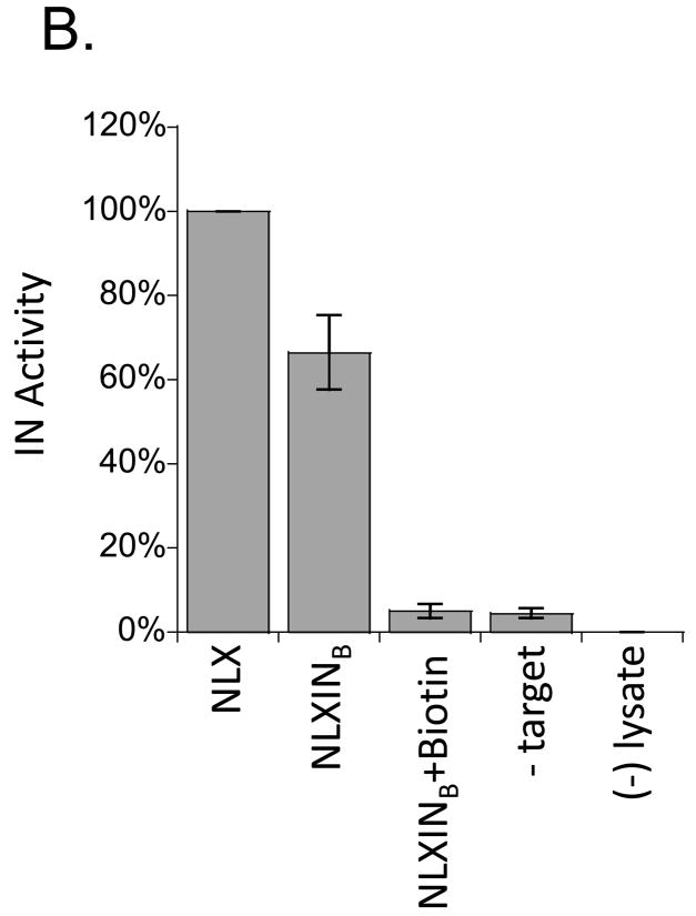
This report describes the adaptation of the biotin ligase BirA-biotin acceptor sequence (BAS) labeling system to biotinylate specific human immunodeficiency virus 1 (HIV-1) proteins in vivo. Two HIV-1 clones were constructed, with the BAS introduced into the matrix region of gag or the integrase region of pol. Specific biotinylation of target proteins in virions was observed when molecular clones were co-expressed with BirA. Both BAS-containing viruses propagated in SupT1 T-cells although replication of the integrase clone was delayed. Further studies demonstrated that the integrase insertion yielded an approximate 40% reduction in single-round infectivity as assessed on MAGI-5 indicator cells, as well as in the in vitro integration activity of preintegration complexes extracted from acutely infected C8166-45 T-cells. Biotinylation of the integrase BAS tag furthermore rendered this virus non-infectious. The matrix viral clone by contrast displayed wild-type behavior under all conditions tested. These results therefore establish a system whereby biotinylated matrix protein in the context of replication-competent virus could be used to label and capture viral protein complexes in vivo.


Figures

Figure 1
Figure 1
Creation of BAS-containing IN and MA molecular clones. (A) The canonical BAS, as described in de Boer et al. (2003). The underlined portion defines the common inserted sequence, as indicated by inverted black triangles in panel B. The asterisk denotes the biotin-acceptor lysine residue within the BAS. (B) BAS placement. The BAS in NLXINB is followed by a stop codon after the C-terminal Ser residue in panel A. NLXMAB harbors the BAS insertion 5 residues N-terminal of the MA-capsid protease cleavage site (indicated by double slash) to permit correct Gag polyprotein processing (Muller et al., 2004). Underlines denote heterologous BAS-abutting linker amino acid residues.
Figure 2
Figure 2
Specific biotinylation of IN and MA in HIV-1 virions. 293T cells were transfected with NLX molecular clones containing the BAS in IN (pNLXINB) or MA (pNLXMAB), alone or with the BirA expression plasmid as indicated. Virion lysates (top three panels) were probed by Western blotting using the indicated reagents. The bottom panel indicates the cellular level of BirA expression.
Figure 3
Figure 3
Construction and characterization of 293T cell lines stably expressing BirA, and SA-capture of biotinylated IN and MA. (A) 293T cells were transfected with pc6BirA and selected using Blasticidin as described in Materials and Methods. Clonal 293T.BirA cell lines were obtained by limiting dilution, expanded, and assayed for BirA expression by Western blotting. (B) SA capture of biotinylated IN and MA proteins from purified virions and cell lysates. Virus was produced by transfection of 293T.BirA cells as described in the legend of Fig. 2. Virus and cell lysates were resuspended in RIPB buffer, a pre-sample removed (Pre), and viral proteins captured with SA-agarose beads (AP). After the beads were pelleted, a post sample was removed (Pt), then the beads were washed extensively with RIPB buffer. Bound proteins were released by boiling in sample buffer, and samples separated by SDS-PAGE were detected using SA-HRP or anti-MA antibody as denoted beneath each blot. Approximate MW sizes are shown to the left of the blots, and viral proteins are indicated on the right.
Figure 3
Figure 3
Construction and characterization of 293T cell lines stably expressing BirA, and SA-capture of biotinylated IN and MA. (A) 293T cells were transfected with pc6BirA and selected using Blasticidin as described in Materials and Methods. Clonal 293T.BirA cell lines were obtained by limiting dilution, expanded, and assayed for BirA expression by Western blotting. (B) SA capture of biotinylated IN and MA proteins from purified virions and cell lysates. Virus was produced by transfection of 293T.BirA cells as described in the legend of Fig. 2. Virus and cell lysates were resuspended in RIPB buffer, a pre-sample removed (Pre), and viral proteins captured with SA-agarose beads (AP). After the beads were pelleted, a post sample was removed (Pt), then the beads were washed extensively with RIPB buffer. Bound proteins were released by boiling in sample buffer, and samples separated by SDS-PAGE were detected using SA-HRP or anti-MA antibody as denoted beneath each blot. Approximate MW sizes are shown to the left of the blots, and viral proteins are indicated on the right.
Figure 4
Figure 4
Replication profiles of BAS-containing viruses. (A) Replication kinetics in SupT1 cells; note the replicating virus is not biotinylated due to the lack of BirA expression in these cells. Results are representative of at least two independent experiments. (B) MAGI-5 cell titer of biotinylated viruses. Virus infectivity was determined after 2 d post-infection by fixing and staining the cells for β-galactosidase expression. Error bars denote the standard deviation from 9 independent infections.
Figure 4
Figure 4
Replication profiles of BAS-containing viruses. (A) Replication kinetics in SupT1 cells; note the replicating virus is not biotinylated due to the lack of BirA expression in these cells. Results are representative of at least two independent experiments. (B) MAGI-5 cell titer of biotinylated viruses. Virus infectivity was determined after 2 d post-infection by fixing and staining the cells for β-galactosidase expression. Error bars denote the standard deviation from 9 independent infections.
Figure 5
Figure 5
NLXINB yields defective PICs. (A) Schematic representation of nested PCR assay used to measure integration activity. The linker primer is represented by a black dashed line, the HIV sequence as a gray line, and target plasmid and primers as solid black lines. (B) PIC activity of NLXINB produced in the absence and presence of IN biotinylation. Samples were prepared in parallel, and results normalized for levels of cDNA synthesis are provided as percentage of activity compared to wild-type HIV-1NLX. No target (-Target) and uninfected lysate ((−) lysate) controls are shown. Data is representative of two independent experiments.
Figure 5
Figure 5
NLXINB yields defective PICs. (A) Schematic representation of nested PCR assay used to measure integration activity. The linker primer is represented by a black dashed line, the HIV sequence as a gray line, and target plasmid and primers as solid black lines. (B) PIC activity of NLXINB produced in the absence and presence of IN biotinylation. Samples were prepared in parallel, and results normalized for levels of cDNA synthesis are provided as percentage of activity compared to wild-type HIV-1NLX. No target (-Target) and uninfected lysate ((−) lysate) controls are shown. Data is representative of two independent experiments.

Similar articles

Cited by

References

    1. Beckett D, Kovaleva E, Schatz P. A minimal peptide substrate in biotin holoenzyme synthetase-catalyzed biotinylation. Protein Sci. 1999;8:921–929. - PMC - PubMed
    1. Belshan M, Mahnke LA, Ratner L. Conserved amino acids of the human immunodeficiency virus type 2 Vpx nuclear localization signal are critical for nuclear targeting of the viral preintegration complex in non-dividing cells. Virology. 2006;346:118–126. - PubMed
    1. Brown HE, Chen H, Engelman A. Structure-based mutagenesis of the human immunodeficiency virus type 1 DNA attachment site: effects on integration and cDNA synthesis. J Virol. 1999;73:9011–9020. - PMC - PubMed
    1. Brussel A, Sonigo P. Analysis of early human immunodeficiency virus type 1 DNA synthesis by use of a new sensitive assay for quantifying integrated provirus. J Virol. 2003;77:10119–10124. - PMC - PubMed
    1. Bukrinsky MI, Sharova N, McDonald TL, Pushkarskaya T, Tarpley WG, Stevenson M. Association of integrase, matrix, and reverse transcriptase antigens of human immunodeficiency virus type 1 with viral nucleic acids following acute infection. Proc Natl Acad Sci USA. 1993;90:6125–6129. - PMC - PubMed

Publication types

MeSH terms

Substances

LinkOut - more resources