. 2009 Jun 12;27(29):3912-20.
doi: 10.1016/j.vaccine.2009.04.001. Epub 2009 Apr 23.

Synthetic peptides coupled to the surface of liposomes effectively induce SARS coronavirus-specific cytotoxic T lymphocytes and viral clearance in HLA-A*0201 transgenic mice

Affiliations

Synthetic peptides coupled to the surface of liposomes effectively induce SARS coronavirus-specific cytotoxic T lymphocytes and viral clearance in HLA-A*0201 transgenic mice

Satoshi Ohno et al. Vaccine. .

Abstract

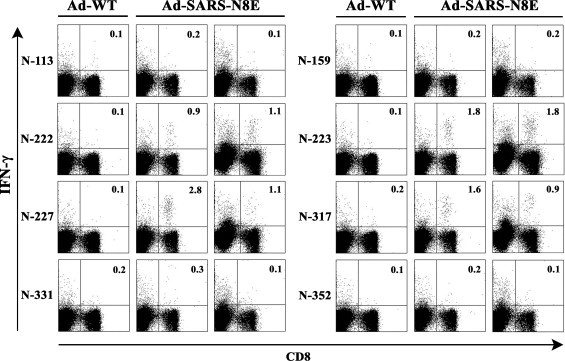
We investigated whether the surface-linked liposomal peptide was applicable to a vaccine based on cytotoxic T lymphocytes (CTLs) against severe acute respiratory syndrome (SARS) coronavirus (SARS-CoV). We first identified four HLA-A*0201-restricted CTL epitopes derived from SARS-CoV using HLA-A*0201 transgenic mice and recombinant adenovirus expressing predicted epitopes. These peptides were coupled to the surface of liposomes, and inoculated into mice. Two of the liposomal peptides were effective for peptide-specific CTL induction, and one of them was efficient for the clearance of vaccinia virus expressing epitopes of SARS-CoV, suggesting that the surface-linked liposomal peptide might offer an effective CTL-based vaccine against SARS.


Figures

Fig. 1
Fig. 1
(A) Nucleotide and amino acid sequences of eight predicted epitopes (N-113, N-159, N-222, N-223, N-227, N-317, N-331 and N-352) with flanking amino acid residues encoded in a minigene, termed SARS-N8E. (B) Expression of the SARS-N8E fusion protein. 293 T cells were infected with either Ad-WT (WT) or Ad-SARS-N8E (N8E). After 2 days’ incubation, cells were lysed and separated on by SDS-12% polyacrylamide gel electrophoresis and subjected to Western blotting analysis with the anti-FLAG antibody. The positions of protein molecular mass markers (in kDa) are shown in the figure, and an arrow indicates the band of the SARS-N8E fusion protein.
Fig. 2
Fig. 2
Intracellular IFN-γ staining of CD8+ T cells specific for SARS-CoV-N-derived peptides in spleen cells of mice immunized with either Ad-SARS-N8E or Ad-WT. HHD mice were immunized i.p. once with either Ad-SARS-N8E or Ad-WT. One week later, spleen cells were prepared and stimulated with each of the eight predicted peptides (N-113, N-159, N-222, N-223, N-227, N-317, N-331, and N-352) for 5 h. Cells were then stained for their surface expression of CD8 (x-axis) and their intracellular expression of IFN-γ (y-axis). The numbers shown indicate the percentages of intracellular IFN-γ+ cells within CD8+ T cell. The data shown are representative of three independent experiments.
Fig. 3
Fig. 3
CTL activities specific for eight predicted epitopes derived from SARS-CoV-N in mice immunized with Ad-SARS-N8E. HHD mice were immunized i.p. with either Ad-SARS-N8E (reverse triangles) or Ad-WT (circles). Two weeks after immunization, spleen cells were prepared and stimulated in vitro with each of the eight predicted epitopes (N-113, N-159, N-222, N-223, N-227, N-317, N-331, and N-352) derived from SARS-CoV N protein. After 1 week, 51Cr-release assays were performed at various E:T ratios with RMA-HHD cells pulsed with (open symbols) or without (solid symbols) a relevant peptide as target. Data are shown as the means ± SD of triplicate wells. The experiment was repeated twice with similar results. At least three mice per group were used in each experiment.
Fig. 4
Fig. 4
In vivo killing of peptide-pulsed target cells in HHD mice immunized with Ad-SARS-N8E. HHD mice were immunized with either Ad-WT or Ad-SARS-N8E. One week later, an equal number of each peptide (N-222, N-223, N-227, or N-317)-pulsed CFSEhigh targets and unpulsed CFSElow targets were transferred into the immunized mice by i.v. injection. After 12 h, CFSE-labeled cells were recovered from spleens of recipient mice and analyzed by flow cytometry. The experiment was repeated three times with similar results. The numbers show the percentages of specific lysis.
Fig. 5
Fig. 5
Intracellular IFN-γ staining of CD8+ T cells specific for SARS-CoV-N-derived peptides in spleen cells of mice immunized with surface-linked liposomal peptides. HHD mice received one injection (1×) of either Lip-N222, Lip-N223, Lip-N227 or Lip-N317, or three injections (3×) of Lip-N317 together with a liposomal helper peptide and CpG. After 1 week, spleen cells were prepared and stimulated with a relevant peptide (N-222, N-223, N-227 or N-317) for 5 h. Cells were then stained for their surface expression of CD8 (x-axis) and their intracellular expression of IFN-γ (y-axis). The numbers shown indicate the percentages of intracellular IFN-γ+ cells within CD8+ T cell. The data shown are representative of three independent experiments.
Fig. 6
Fig. 6
Cross reactivity between N-222 and N-223 peptides. HHD mice were immunized with either Lip-N222 or Lip-N-223 together with a liposomal helper peptide and CpG in the footpad. One week later, spleen cells were prepared and stimulated in vitro with or without either N-222 or N-223 for 5 h. Cells were then stained for their surface expression of CD8 (x-axis) and their intracellular expression of IFN-γ (y-axis). The numbers shown indicate the percentages of intracellular IFN-γ+ cells within CD8+ T cell. The experiment was repeated twice with similar results.
Fig. 7
Fig. 7
In vivo killing activities specific for N-223 and N-227 in HHD mice immunized with surface-linked liposomal peptides. HHD mice were immunized once with either Lip-N223 or Lip-N227 together with a liposomal helper peptide and CpG in the footpad. One week later, an equal number of a relevant peptide (N-223 or N-227)-pulsed CFSEhigh targets and unpulsed CFSElow targets were transferred into the immunized mice by i.v. injection. After 12 h, CFSE-labeled cells were recovered from spleens of recipient mice and analyzed by flow cytometry. The numbers show the percentages of specific lysis. The experiment was repeated twice.
Fig. 8
Fig. 8
Resistance to infection with vaccinia virus expressing N-223 in mice immunized with Lip-N223. HHD mice were immunized twice with either Lip-N223 or empty liposomes (Empty-Lip) along with a liposomal helper peptide and CpG at 2-week intervals. Two weeks later, mice were challenged i.p. with 1 × 106 PFU of either VV-SARS-N8E (VV-SARS) or VV-WT. Mice were then sacrificed 5 days after challenge, and viral titers in the ovaries were measured. All titrations were performed in duplicates, and the average PFU per mouse is shown in the figure. Three mice were used in each group, and data are shown as the mean ± SD of three mice. *P < 0.01.

Similar articles

Cited by

References

    1. Groneberg D.A., Zhang L., Welte T., Zabel P., Chung K.F. Severe acute respiratory syndrome: global initiatives for disease diagnosis. Q J Med. 2003;96(11):845–852. - PMC - PubMed
    1. Drosten C., Gunther S., Preiser W., van der Werf S., Brodt H.R., Becker S. Identification of a novel coronavirus in patients with severe acute respiratory syndrome. N Engl J Med. 2003;348(20):1967–1976. - PubMed
    1. Ksiazek T.G., Erdman D., Goldsmith C.S., Zaki S.R., Peret T., Emery S. A novel coronavirus associated with severe acute respiratory syndrome. N Engl J Med. 2003;348(20):1953–1966. - PubMed
    1. Peiris J.S.M., Lai S.T., Poon L.L.M., Guan Y., Yam L.Y.C., Lim W. Coronavirus as a possible cause of severe acute respiratory syndrome. Lancet. 2003;361(9371):1319–1325. - PMC - PubMed
    1. Marra M.A., Jones S.J.M., Astell C.R., Holt R.A., Brooks-Wilson A., Butterfield Y.S.N. The genome sequence of the SARS-associated coronavirus. Science. 2003;300(5624):1399–1404. - PubMed

Publication types

MeSH terms