. 2009 Jul 31;284(31):20909-16.
doi: 10.1074/jbc.M109.014472. Epub 2009 Jun 2.

Phosphorylated Tau interacts with c-Jun N-terminal kinase-interacting protein 1 (JIP1) in Alzheimer disease

Affiliations

Phosphorylated Tau interacts with c-Jun N-terminal kinase-interacting protein 1 (JIP1) in Alzheimer disease

Lars M Ittner et al. J Biol Chem. .

Abstract

In Alzheimer disease (AD) and frontotemporal dementia the microtubule-associated protein Tau becomes progressively hyperphosphorylated, eventually forming aggregates. However, how Tau dysfunction is associated with functional impairment is only partly understood, especially at early stages when Tau is mislocalized but has not yet formed aggregates. Impaired axonal transport has been proposed as a potential pathomechanism, based on cellular Tau models and Tau transgenic mice. We recently reported K369I mutant Tau transgenic K3 mice with axonal transport defects that suggested a cargo-selective impairment of kinesin-driven anterograde transport by Tau. Here, we show that kinesin motor complex formation is disturbed in the K3 mice. We show that under pathological conditions hyperphosphorylated Tau interacts with c-Jun N-terminal kinase- interacting protein 1 (JIP1), which is associated with the kinesin motor protein complex. As a result, transport of JIP1 into the axon is impaired, causing JIP1 to accumulate in the cell body. Because we found trapping of JIP1 and a pathological Tau/JIP1 interaction also in AD brain, this may have pathomechanistic implications in diseases with a Tau pathology. This is supported by JIP1 sequestration in the cell body of Tau-transfected primary neuronal cultures. The pathological Tau/JIP1 interaction requires phosphorylation of Tau, and Tau competes with the physiological binding of JIP1 to kinesin light chain. Because JIP1 is involved in regulating cargo binding to kinesin motors, our findings may, at least in part, explain how hyperphosphorylated Tau mediates impaired axonal transport in AD and frontotemporal dementia.


Figures

FIGURE 1.
FIGURE 1.
Disturbed interaction of JIP1 with the kinesin-I motor complex in K3 mice. A, K369I mutant human Tau expressing K3 mice (HT7) phosphorylate Tau at multiple sites, including AT8, AT180, PHF-1, and AT270, as shown for 4-month-old mice compared with wild-type (wt). Despite axonal transport defects in K3 mice (17), protein levels of the motor proteins kinesin heavy chain (KIF5B), KLC, and JIP1 are not altered. GAPDH served as control for equal loading. Brain extracts from four different mice per group were analyzed. B, although an intact JIP1·KLC·KIF5B complex could be immunoprecipitated from wild-type mouse brain extracts with either an antibody against JIP1, KLC, or KIF5B, in K3 brain only the KLC/KIF5B interaction could be revealed, with no JIP1 in the complex. Representative blots from at least three independent experiments are shown. C, IP of Tau from K3 and wild-type brain extracts reveals a Tau/JIP1 interaction in K3 and not wild-type brain. Similarly, IP with a JIP1 antibody co-precipitates Tau from K3 but not wild-type mouse brain. Representative blots from three experiments are shown.
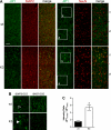
FIGURE 2.
FIGURE 2.
Tau-dependent re-distribution of JIP1 in K3 neurons. A, immunohistochemistry of sagittal brain sections reveals a predominantly axonal staining of JIP1 (green) in wild-type mice, with little staining of neuronal cell bodies. In contrast, JIP1 accumulates in the cell body of cortical neurons of K3 mice, whereas axons hardly contain JIP1. Neurons were counter-stained with microtubule-associated protein 2 (MAP2) or neuronal nuclei (NeuN) (red). Scale bar, 100 μm. B, higher magnification (boxes in A) of layer V neuronal cell bodies (soma (s)) and their axons (a) passing through neuronal layer VI. JIP1 accumulates in K3 neuronal cell bodies (arrowhead), whereas they are hardly stainable in wild-type (wt) neurons. C, thus, the ratio of somatic versus axonal JIP1 is significantly increased in K3 mice, indicating trapping of JIP1 in the cell body (*, p < 0.0001).
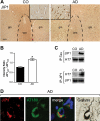
FIGURE 3.
FIGURE 3.
Pathological re-distribution of JIP1 in AD brain. A, low magnification images of JIP1-stained (brown) sections from the temporal cortex of control (CO) brains show a low signal in gray matter (gm) (inset at higher magnification) and intensive staining of axons in the white matter (wm). In contrast, the white matter of AD tissue sections stains less for JIP1, whereas gray matter neurons (inset) show intensive JIP1 staining. Scale bar, 250 μm. B, quantification reveals a re-distribution of JIP1 from the axon to the soma, as shown by an increased ratio of somatic versus axonal staining (*, p < 0.0001). C, JIP1 co-immunoprecipitates with Tau from human AD but not control (CO) brain extracts. Similarly, IP using a JIP1 antibody co-precipitates Tau from AD but not CO brains. Representative blots from three experiments are shown. D, JIP1 (red)/AT180 (green) co-immunofluorescence staining shows co-localization (merge) in NFTs, which stain with Gallyas silver.
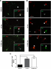
FIGURE 4.
FIGURE 4.
Impaired axonal distribution of JIP1 is not caused by the K369I mutation of Tau. A, to determine whether the impaired axonal localization of JIP1 is due to the presence of K369I mutant Tau rather than elevated levels of hyperphosphorylated Tau per se, primary hippocampal neurons were transfected with MOCK, V5-wt-Tau, or V5-K369I mutant Tau together with green fluorescent protein (GFP) to visualize axonal tracing. In MOCK co-transfected neurons, JIP1 undergoes axonal transport and accumulates in growth cones (arrowheads; inset). In contrast, neurons co-transfected with V5-Tau (yellow merge) fail to accumulate JIP1 in growth cones (open arrowheads; inset). Note the intense JIP1 staining of the growth cone in untransfected neurons (arrowhead). Co-transfection of V5-K369I mutant Tau also results in a decreased JIP1 staining of growth cones. Scale bar, 50 μm. wt, wild type. B, co-transfection of primary hippocampal neurons with either V5-wt-Tau or V5-K369I mutant Tau together with Myc-JIP1 results in intensive JIP1 staining of the cell body (arrowheads), that is not found when the neurons are only transfected with the Myc-JIP1 construct (open arrowhead). Inset, staining for Tau. Scale bar, 50 μm. C, when staining is quantified, Myc-JIP1 fluorescence intensity shows increased levels of JIP1 in neurons that co-express either V5-wt-Tau or V5-K369I mutant Tau compared with co-transfection with an empty plasmid (MOCK; *, p < 0.0001). Thus, an increase of Tau in the cell body of neurons results in retention of JIP1, along with a decreased axonal distribution.
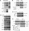
FIGURE 5.
FIGURE 5.
Hyperphosphorylation of Tau is required for Tau to compete with KLC in the interaction with JIP1. A, recombinant hyperphosphorylated Tau carrying a carboxyl-terminal V5 and six-histidine tag (P-Tau-6×His) was used as bait to identify interaction partners of the kinesin-I-JIP complex from wild-type mouse brain extracts (load). JIP1 (asterisk) was pulled down with P-Tau-His6. Neither JIP2, JIP3, KLC, nor kinesin heavy chain (KIF5B) co-precipitated with P-Tau-His6. Recombinant Tau was visualized with V5 and Tau-5 antibodies. B, COS7 cells were co-transfected with Myc-JIP1 and V5-Tau (lane 1) or V5-KLC (lane 2). Immunoprecipitation with a Myc antibody precipitates JIP1 and co-precipitates V5-KLC but not V5-Tau. GAPDH served as loading control. C, treatment of COS7 cells that have been co-transfected with Myc-JIP1 and V5-Tau with increasing concentrations of the protein phosphatase inhibitor OA causes increased phosphorylation of Tau at multiple sites including AT8, AT180, PHF-1, and AT270. OA treatment did not affect Tau levels, as revealed by Tau-1. GAPDH served as control for equal loading. D, co-immunoprecipitation from OA-treated transfected cells (d) results in a dose-dependent increase of V5-Tau co-precipitated with Myc-JIP1. E, COS7 cells transfected with Myc-JIP1 and V5-KLC with (lanes 1 and 3) and without V5-Tau (lane 2) treated with 20 nm OA. OA impedes co-precipitation of V5-KLC with Myc-JIP1, most significantly in the presence of V5-Tau that then co-precipitates with Myc-JIP1. Quantification is shown for three independent experiments normalized for levels in the absence of OA (lane 1; *, p < 0.001).

Similar articles

Cited by

  • Lessons from tau-deficient mice.
    Ke YD, Suchowerska AK, van der Hoven J, De Silva DM, Wu CW, van Eersel J, Ittner A, Ittner LM. Ke YD, et al. Int J Alzheimers Dis. 2012;2012:873270. doi: 10.1155/2012/873270. Epub 2012 Jun 6. Int J Alzheimers Dis. 2012. PMID: 22720190 Free PMC article.
  • Yeast as a model system to study tau biology.
    De Vos A, Anandhakumar J, Van den Brande J, Verduyckt M, Franssens V, Winderickx J, Swinnen E. De Vos A, et al. Int J Alzheimers Dis. 2011 Apr 6;2011:428970. doi: 10.4061/2011/428970. Int J Alzheimers Dis. 2011. PMID: 21559193 Free PMC article.
  • MSUT2 regulates tau spreading via adenosinergic signaling mediated ASAP1 pathway in neurons.
    Xu H, Qiu Q, Hu P, Hoxha K, Jang E, O'Reilly M, Kim C, He Z, Marotta N, Changolkar L, Zhang B, Wu H, Schellenberg GD, Kraemer B, Luk KC, Lee EB, Trojanowski JQ, Brunden KR, Lee VM. Xu H, et al. Acta Neuropathol. 2024 Mar 12;147(1):55. doi: 10.1007/s00401-024-02703-3. Acta Neuropathol. 2024. PMID: 38472475 Free PMC article.
  • The role of tau kinases in Alzheimer's disease.
    Dolan PJ, Johnson GV. Dolan PJ, et al. Curr Opin Drug Discov Devel. 2010 Sep;13(5):595-603. Curr Opin Drug Discov Devel. 2010. PMID: 20812151 Free PMC article. Review.
  • Tau as a potential therapeutic target for ischemic stroke.
    Chen X, Jiang H. Chen X, et al. Aging (Albany NY). 2019 Dec 16;11(24):12827-12843. doi: 10.18632/aging.102547. Epub 2019 Dec 16. Aging (Albany NY). 2019. PMID: 31841442 Free PMC article. Review.

References

    1. Lee V. M., Goedert M., Trojanowski J. Q. (2001) Annu. Rev. Neurosci. 24, 1121–1159 - PubMed
    1. Goedert M., Spillantini M. G., Jakes R., Rutherford D., Crowther R. A. (1989) Neuron 3, 519–526 - PubMed
    1. Goedert M., Wischik C. M., Crowther R. A., Walker J. E., Klug A. (1988) Proc. Natl. Acad. Sci. U.S.A. 85, 4051–4055 - PMC - PubMed
    1. Alonso A. C., Grundke-Iqbal I., Iqbal K. (1996) Nat Med 2, 783–787 - PubMed
    1. Chen F., David D., Ferrari A., Götz J. (2004) Curr. Drug Targets 5, 503–515 - PubMed

Publication types