. 2009 Aug 7;284(32):21265-9.
doi: 10.1074/jbc.M109.019760. Epub 2009 Jun 1.

Helix straightening as an activation mechanism in the gelsolin superfamily of actin regulatory proteins

Affiliations

Helix straightening as an activation mechanism in the gelsolin superfamily of actin regulatory proteins

Hui Wang et al. J Biol Chem. .

Abstract

Villin and gelsolin consist of six homologous domains of the gelsolin/cofilin fold (V1-V6 and G1-G6, respectively). Villin differs from gelsolin in possessing at its C terminus an unrelated seventh domain, the villin headpiece. Here, we present the crystal structure of villin domain V6 in an environment in which intact villin would be inactive, in the absence of bound Ca(2+) or phosphorylation. The structure of V6 more closely resembles that of the activated form of G6, which contains one bound Ca(2+), rather than that of the calcium ion-free form of G6 within intact inactive gelsolin. Strikingly apparent is that the long helix in V6 is straight, as found in the activated form of G6, as opposed to the kinked version in inactive gelsolin. Molecular dynamics calculations suggest that the preferable conformation for this helix in the isolated G6 domain is also straight in the absence of Ca(2+) and other gelsolin domains. However, the G6 helix bends in intact calcium ion-free gelsolin to allow interaction with G2 and G4. We suggest that a similar situation exists in villin. Within the intact protein, a bent V6 helix, when triggered by Ca(2+), straightens and helps push apart adjacent domains to expose actin-binding sites within the protein. The sixth domain in this superfamily of proteins serves as a keystone that locks together a compact ensemble of domains in an inactive state. Perturbing the keystone initiates reorganization of the structure to reveal previously buried actin-binding sites.


Figures

FIGURE 1.
FIGURE 1.
Structural comparison of villin V6 (Ca2+-free) with gelsolin G6 (Ca2+-bound and Ca2+-free). A, schematic representation of isolated Ca2+-free V6. B, key residues that are involved in the putative V6 calcium-binding site, viewed from below with respect to A. C and D, Ca2+-bound form of gelsolin G6, displaying a straight helix, taken from the Ca2+-bound form of G4–G6 (Protein Data Bank code 1p8x). E and F, Ca2+-free form of G6 revealing a kinked helix and a translocated AB loop, taken from the structure of whole plasma gelsolin (Protein Data Bank code 1d0n). The Ca2+-binding residues are dislocated in the absence of Ca2+ compared with D. A hydrogen bond between Asn647 and Arg702 links the bending of the helix to movement of the AB loop. Protein representations were generated here and in the figures that follow using PyMOL.
FIGURE 2.
FIGURE 2.
Comparison of the domain 4 interaction surfaces of G6 and V6. A, representation of Ca2+-free gelsolin showing the central position of G6 within the inactive structure. The black sphere pinpoints the unoccupied Ca2+-binding site within G6. B, surface charge representation of G6 from A, rotated ∼90° around the vertical axis, showing the interaction surface with G4 via a common β-sheet. C and D, Ca2+-bound G6 (C) and Ca2+-free V6 (D) superimposed on G6 in B. These models show minor steric clashes with the G4 loop that follows the β-sheet interaction in B while the alternating charge distribution at the β-strand edge is maintained.
FIGURE 3.
FIGURE 3.
Comparison of the domain 2 interaction surfaces of G6 and V6. A, Arg168 and Arg169, from G2, bind to Asp670 and a negatively charged surface on G6 in the Ca2+-free form of gelsolin. The AB loop is distant from G2 in this conformation. B, Ca2+ binding by G6 involves direct coordination with Asp670. Superposition of Ca2+-bound G6 on A reveals that the AB loop moves to clash with G2 in this model. C, superposition of Ca2+-free V6 on A creates a model that demonstrates that Asp648 is similarly placed to activate villin via Ca2+ binding or to secure the Ca2+-free structure through binding to V2. In this model, the AB loop clashes with domain 2 and, as such, would have to move to bind V2.
FIGURE 4.
FIGURE 4.
Domain 6 interactions with actin. A, the structure of G4–G6·actin (Protein Data Bank code 1h1v) shows that the actin helix comprising residues 307–321 contacts the AB loop of G6. B, surface charge representation of Ca2+-bound G6 shows the interaction with actin residues 307–321. Ca2+-bound G6 is oriented as in Fig. 1C. C, superposition of Ca2+-free V6 onto B produces a model that suggests that V6 might retain the ability to interact with these actin residues. D, similar modeling of the transformation of G6 to a Ca2+-free state disturbs that interaction.
FIGURE 5.
FIGURE 5.
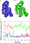
Straightening of the G6 helix in isolation. A, superposition of backbone configurations in 10 ns of a molecular dynamics trajectory in which the initial structure was the active form of G6 (from Protein Data Bank code 1h1v). B, superposition of backbone configurations in the last 10 ns (after relaxation to the straightened helix conformation) of a molecular dynamics trajectory, shown in C, in which the initial structure was the inactive form of G6 (from Protein Data Bank code 1d0n). C, fit of the long helix to the initial, bent conformation (green) and to the straightened helix (from Protein Data Bank code 1h1v) (red) over a 15-ns molecular dynamics simulation that started with the conformation from Protein Data Bank code 1d0n. As a control, the fit to the straight helix (from Protein Data Bank code 1h1v) is also shown for a simulation starting from the same structure (blue). In this trajectory, produced in the absence of other gelsolin domains and Ca2+, the long helix relaxes toward the crystallographic structure of activated G6 (from Protein Data Bank code 1h1v) in less than 10 ns. RMSD, root mean square deviation.

Similar articles

Cited by

References

    1. dos Remedios C. G., Chhabra D., Kekic M., Dedova I. V., Tsubakihara M., Berry D. A., Nosworthy N. J. (2003) Physiol. Rev. 83, 433–473 - PubMed
    1. McGough A. M., Staiger C. J., Min J. K., Simonetti K. D. (2003) FEBS Lett. 552, 75–81 - PubMed
    1. Khurana S., George S. P. (2008) FEBS Lett. 582, 2128–2139 - PMC - PubMed
    1. Janmey P. A., Matsudaira P. T. (1988) J. Biol. Chem. 263, 16738–16743 - PubMed
    1. George S. P., Wang Y., Mathew S., Srinivasan K., Khurana S. (2007) J. Biol. Chem. 282, 26528–26541 - PubMed

Publication types