. 2009 Jul 15;587(Pt 14):3539-59.
doi: 10.1113/jphysiol.2008.167502. Epub 2009 Jun 2.

Abdominal expiratory activity in the rat brainstem-spinal cord in situ: patterns, origins and implications for respiratory rhythm generation

Affiliations

Abdominal expiratory activity in the rat brainstem-spinal cord in situ: patterns, origins and implications for respiratory rhythm generation

A P L Abdala et al. J Physiol. .

Abstract

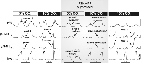
We studied respiratory neural activity generated during expiration. Motoneuronal activity was recorded simultaneously from abdominal (AbN), phrenic (PN), hypoglossal (HN) and central vagus nerves from neonatal and juvenile rats in situ. During eupnoeic activity, low-amplitude post-inspiratory (post-I) discharge was only present in AbN motor outflow. Expression of AbN late-expiratory (late-E) activity, preceding PN bursts, occurred during hypercapnia. Biphasic expiratory (biphasic-E) activity with pre-inspiratory (pre-I) and post-I discharges occurred only during eucapnic anoxia or hypercapnic anoxia. Late-E activity generated during hypercapnia (7-10% CO(2)) was abolished with pontine transections or chemical suppression of retrotrapezoid nucleus/ventrolateral parafacial (RTN/vlPF). AbN late-E activity during hypercapnia is coupled with augmented pre-I discharge in HN, truncated PN burst, and was quiescent during inspiration. Our data suggest that the pons provides a necessary excitatory drive to an additional neural oscillatory mechanism that is only activated under conditions of high respiratory drive to generate late-E activity destined for AbN motoneurones. This mechanism may arise from neurons located in the RTN/vlPF or the latter may relay late-E activity generated elsewhere. We hypothesize that this oscillatory mechanism is not a necessary component of the respiratory central pattern generator but constitutes a defensive mechanism activated under critical metabolic conditions to provide forced expiration and reduced upper airway resistance simultaneously. Possible interactions of this oscillator with components of the brainstem respiratory network are discussed.


Figures

Figure 2
Figure 2. Characteristics of the respiratory abdominal motor outflow in juvenile and neonatal rat in situ preparations
Representative activity patterns of phrenic (PN), hypoglossal (HN), lumbar abdominal (AbN) and central vagus (cVN) nerves recorded simultaneously during eupnoea (5% CO2) and hypercapnia (7%, 10% CO2) in juvenile (A) and neonatal rats (B). Raw (grey traces) and integrated (black traces) motor nerve outputs; vertical dashed lines indicate onset of HN bursts. Hypercapnia (7% CO2) elicited typical AbN late-E bursts (arrowheads) that exhibited cycle-to-cycle ‘skipping’ but were stable at 10% CO2. Insets in A: late-E AbN bursts were correlated with shorter duration of the preceding cVN post-inspiratory activity (see also Fig. 3E). The time scale in the insets indicates time from the end of inspiration in PN.
Figure 3
Figure 3. Effect of abdominal late expiratory discharge on phrenic activity
Graphs show phrenic nerve (PN) burst rate (A), burst duration (TI) (B), inter-burst duration (TE) (D) and amplitude (E) under eupnoea (5% CO2) and hypercapnia (7–10% CO2). We averaged cycles without (grey bars) and with abdominal (AbN) late-E activity (black bars). All values represent mean ±s.e.m. (error bars), n= 10. The representative cycle-triggered average (CTA) shows PN bursts not preceded by late-E AbN activity (C) under hypercapnia (7% CO2). CTA in C represents 30 cycles not preceded by late-E AbN bursts. The presence of AbN (grey) late-E bursts correlated with shortened duration of succeeding PN bursts (black) and prolonged the PN inter-burst interval without changing its amplitude (F). CTA in F is an average of 20 cycles preceded by late-E AbN bursts. Time 0 indicates onset of inspiration on PN.
Figure 1
Figure 1. Abdominal motor activity patterns during high central respiratory drive
Effect of increased respiratory drive on integrated phrenic (PN), hypoglossal (HN), lumbar abdominal (AbN) and central vagus (cVN) outflows recorded simultaneously. A, during eupnoea in the juvenile rat (a1) AbN activity exhibits low amplitude post-I discharge. The response to anoxia (95% N2 and 5% CO2) was divided into 3 phases: inspiratory excitation (a2), inspiratory depression (a3) and gasping (a4). During phases a2 and a3 augmenting AbN discharge emerged with variable amplitudes. The pattern transformed into biphasic-E AbN activity during gasping (phase a4). Note that the appearance of a single doublet inspiratory burst (third burst prior to a4) was predicted by the model of Wittmeier et al. (2008). B, neonatal rats (n= 3) presented similar motor outflows during gasping with biphasic-E AbN discharges. This activity continued longer (in comparison with juvenile rats) without suffering respiratory arrest. C, bolus injections of NaCN (100 μl, 0.03%) into the perfusate to activate peripheral chemoreceptors also induced biphasic-E activity in the AbN in juvenile rats.
Figure 4
Figure 4. Occurrence of late-E abdominal (AbN) activity is correlated with alterations of cranial respiratory motor outflow patterns
At 7% CO2, late-E AbN bursts were followed by increased amplitude (A) and advanced onset (B) of HN bursts. Black bars show cycles that were preceded by late-E AbN and grey bars, cycles that were not. C, representative CTAs show an average of 26 HN bursts (black) that were preceded by late-E AbN bursts (grey), and 24 cycles that were not. Time 0 marks onset of inspiration on PN. At 7% CO2, late-E AbN bursts correlated with advanced onset (D) of following cVN burst and advanced offset of post-inspiratory (post-I) activities (E) of the preceding cVN bursts. F, representative CTAs were compiled from 20 cVN bursts (black) that were preceded by late-E AbN bursts (grey) and 30 that were not. All values represent mean ±s.e.m. (error bars).
Figure 5
Figure 5
A, schematic diagram depicting the spatial arrangement of the ventral respiratory column viewed sagittally. We performed precise microtransections of the brainstem indicated by the vertical dashed lines (see Methods). B, activity patterns of phrenic (PN), spinal abdominal (AbN) and central vagus (cVN) nerves from an intact preparation (3-phase pattern) during eucapnia (5% CO2) and hypercapnia (7% CO2, n= 15), and after a ponto-medullary transection. This resulted in a 2-phase pattern in which late-E abdominal bursts are absent during hypercapnia (8.5% CO2). AbN motor output displayed tonic activity throughout expiration, which decremented slightly in the late-E phase (n= 8). After a transection at the rostral boundary of the pre-BötC, a 1-phase pattern was evoked and all expiratory motor output was absent (n= 8). Abbreviations: AmbC: compact nucleus ambiguus; BötC: Bötzinger complex; LRt: lateral reticular nucleus; Pn: pontine nucleus; pre-BötC: pre- Bötzinger complex; RTN/vlPF: retrotrapezoid nucleus and ventrolateral parafacial regions; rVRG: rostral ventral respiratory group; V: trigeminal motor nucleus; VII: facial motor nucleus.
Figure 7
Figure 7
A, we microinjected dye (Pontamine sky blue) after experiments to allow a topographical mapping of the microinjection sites into the retrotrapezoid/ventrolateral parafacial regions (RTN/vlPF). Microinjection centres located in the caudal half of the facial nucleus (VII) and aligned at the medial lateral level of the compact division of nucleus ambiguous (AmbC) produced the most potent inhibition of expiratory motor outflow (both post-I and late-E). B, example of a sagittal section (40 μm, neutral red) of a brainstem at the level of facial nucleus (VII) and the Bötzinger complex (BötC) with a stained microinjection site (arrow); the schematic diagram depicts the outline of the relevant respiratory-related regions. Other abbreviations: Amb: nucleus ambiguus; pre-BötC: pre-Bötzinger complex; XII: hypoglossal nerve rootlets.
Figure 6
Figure 6. The retrotrapezoid/ventrolateral parafacial regions (RTN/vlPF) are relevant for the 3-phase respiratory rhythm and generation of late-E abdominal (AbN) activity
Hypercapnia (10% CO2) induces late-E bursts in the AbN (T12–L1) (left). Bilateral microinjections (n= 11) of isoguvacine hydrochloride (GABAA receptor agonist) to suppress the RTN/vlPF region disrupted the 3-phase respiratory rhythm during eucapnia (5% CO2). Post-inspiratory motor output on both AbN and central vagus (cVN) nerves was reduced and phrenic nerve pattern (PN) was transformed to a ‘square-wave’ shape. RTN/vlPF suppression also abolished late-E AbN bursts during hypercapnia (10% CO2). Late-E activity recovered after isoguvacine was washed out (∼1 h) (right panel). All traces show integrated nerve activities.
Figure 8
Figure 8. Suppression of RTN/vlPF does not affect BötC augmenting E activity but abolishes late-E abdominal discharge
To assess the relevance of the Bötzinger complex (BötC) for late-E abdominal activity, we recorded augmenting expiratory (aug-E) population activity in the BötC simultaneously with phrenic (PN), abdominal (AbN) and central vagus (cVN) nerves as we inhibited the RTN/vlPF region (A). Bilateral microinjections of a GABAA agonist into the RTN/vlPF region abolished AbN late-E bursts completely, but central aug-E activity persisted in the BötC. CTA of integrated BötC population activities under 7% CO2 in cycles that do not contain late-E AbN bursts (B), containing late-E bursts (C) and after RTN/vlPF was suppressed (D). Time 0 indicates beginning of PN inspiration and grey area indicates duration of inspiration. E, maximum firing frequency (% of control at 5% CO2) of BötC augmenting E population before and after RTN/vlPF inhibition. Note that maximum firing frequency of BötC population was increased in association with late-E AbN bursts, and both were abolished by RTN/vlPF suppression, but baseline BötC activity persisted.
Figure 9
Figure 9. Examples of two types of neurones recorded in the same region of the RTN/vlPF during hypercapnia
Traces show integrated phrenic (PN), spinal abdominal (AbN), central vagus (cVN) nerve activities and neurone recorded in RTN/vlPF region with firing rate histograms. A, an example of a rhythmically active late-E neuron. Under hypercapnia (7% CO2), these cells (n= 7 from 5 juvenile rats) fired exclusively in phase with late-E abdominal bursts, but were quiescent in the absence of late-E AbN bursts as in eucapnia (5% CO2). B, example of a RTN CO2-sensitive tonic neurone reported previously (Fortuna et al. 2008) which was intermingled with the neurone in A.
Figure 10
Figure 10. Extended model of the brainstem respiratory network
(See detailed description in Smith et al. 2007.) A, schematic diagram of the extended model showing interactions between specific populations of respiratory neurones within major brainstem respiratory compartments (pons, RTN/vlPF, BötC, pre-BötC, rVRG and cVRG). Each population (shown as a sphere) consists of 50 neurons described in the Hodgkin–Huxley style. This model incorporates an additional late-E population in the RTN/vlPF. This population is identical to the pre-I/I population in the pre-BötC and consists of 50 neurones containing persistent sodium current and interconnected with mutually excitatory connections (Smith et al. 2007). Most connections are based on previously published experimental data (for references see Smith et al. 2007) and some are suggested (ibid). The model includes proposed interactions between the late-E neuronal population in the RTN/vlPF, activated by hypercapnia, and other populations of respiratory neurones. Interactions between late-E and pre-I/I populations are similar to those proposed by Wittmeier et al. (2008). B and Ca–e show activity of selected neuronal populations in the model (B) and motor outputs (AbN, HN, PN in Ca–e). Activity of each population is represented by a histogram of average neuronal spiking frequency within the population (spikes s–1 neurone–1, bin size 30 ms). Hypercapnia was simulated by an additional ‘hypercapnia-evoked’ drive to the late-E population of RTN/vlPF. In B and Cb, the value of this drive was 0.27 (arbitrary units). In Ca–d/e, this drive progressively increased (indicated above each diagram). The late-E activity was expressed and increased frequency with the increase of this drive (Ca–c). Increasing drive above 0.35 converted the late-E pattern in the late-E population and AbN output to biphasic-E, containing a rebound post-I component (Cd, see also Wittmeier et al. 2008). Also similar to the Wittmeier et al. model, suppression of the pre-I/I population in the pre-BötC (by direct inhibition) to simulate the effects of opioids (Janczewski & Feldman, 2006a) eliminates inspiratory inhibition of the late-E population by the early-I population of pre-BötC, and converts the biphasic-E bursts in AbN to prolonged monophasic discharges lacking the rebound post-I component (Ce).

References

    1. Abraham KA, Feingold H, Fuller DD, Jenkins M, Mateika JH, Fregosi RF. Respiratory-related activation of human abdominal muscles during exercise. J Physiol. 2002;541:653–663. - PMC - PubMed
    1. Ballanyi K, Onimaru H, Homma I. Respiratory network function in the isolated brainstem–spinal cord of newborn rats. Prog Neurobiol. 1999;59:583–634. - PubMed
    1. Ballanyi K, Volker A, Richter DW. Anoxia induced functional inactivation of neonatal respiratory neurones in vitro. Neuroreport. 1994;6:165–168. - PubMed
    1. Cohen MI. Neurogenesis of respiratory rhythm in the mammal. Physiol Rev. 1979;59:1105–1173. - PubMed
    1. Connelly CA, Ellenberger HH, Feldman JL. Respiratory activity in retrotrapezoid nucleus in cat. Am J Physiol Lung Cell Mol Physiol. 1990;258:L33–L44. - PubMed

Publication types

LinkOut - more resources