. 2009 Aug 1;587(Pt 15):3955-65.
doi: 10.1113/jphysiol.2009.173302. Epub 2009 Jun 2.

KCNQ1 is the luminal K+ recycling channel during stimulation of gastric acid secretion

Affiliations

KCNQ1 is the luminal K+ recycling channel during stimulation of gastric acid secretion

Penghong Song et al. J Physiol. .

Abstract

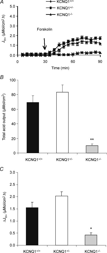
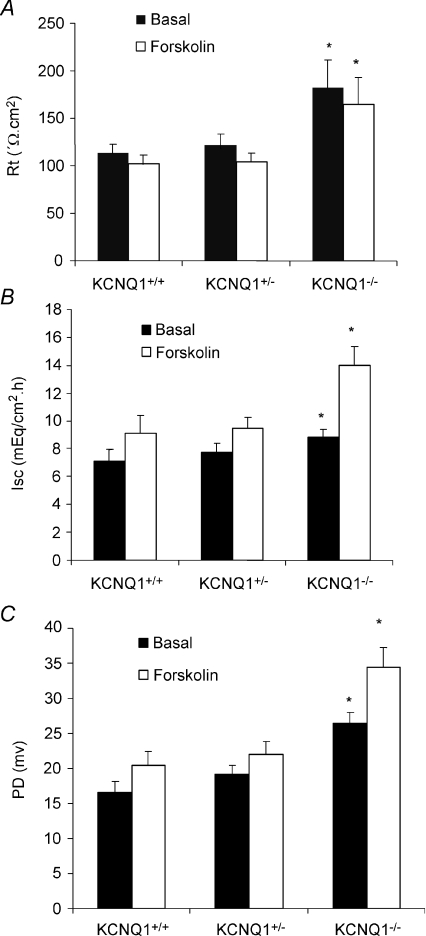
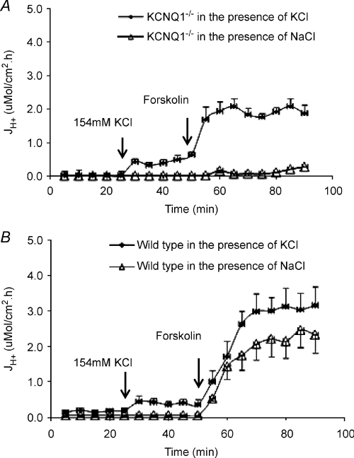
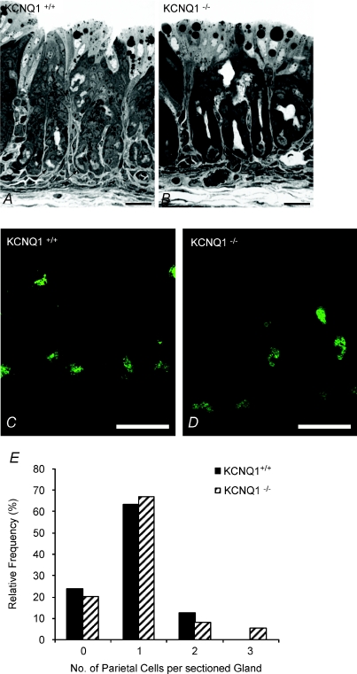
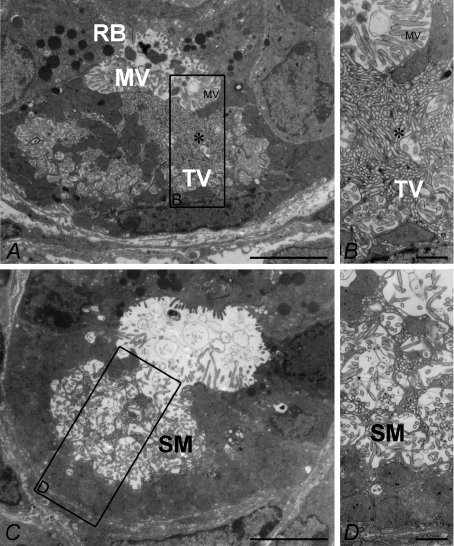
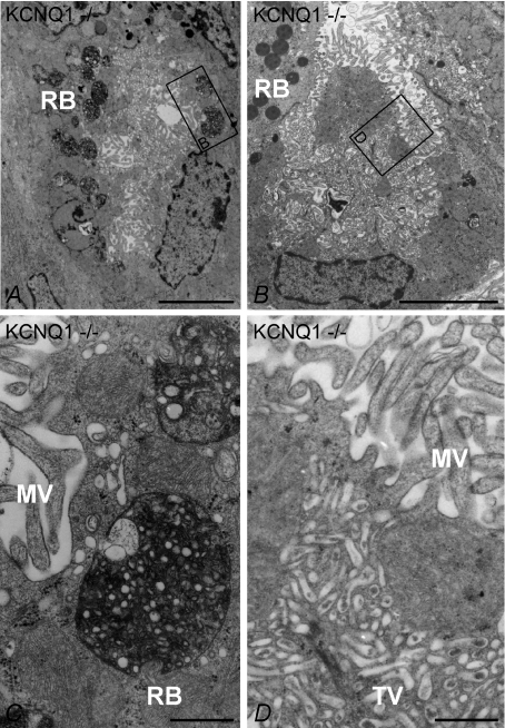
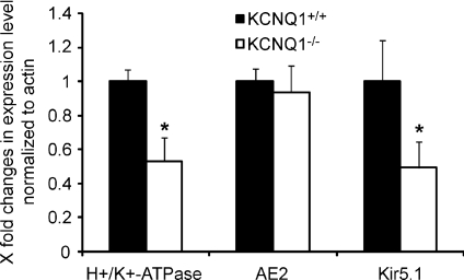
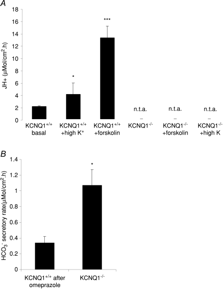
Parietal cell (PC) proton secretion via H(+)/K(+)-ATPase requires apical K(+) recycling. A variety of K(+) channels and transporters are expressed in the PC and the molecular nature of the apical K(+) recycling channel is under debate. This study was undertaken to delineate the exact function of KCNQ1 channels in gastric acid secretion. Acid secretory rates and electrophysiological parameters were determined in gastric mucosae of 7- to 8-day-old KCNQ1(+/+), (+/-) and (-/-) mice. Parietal cell ultrastructure, abundance and gene expression levels were quantified. Glandular structure and PC abundance, and housekeeping gene expression did not differ between the KCNQ1(-/-) and (+/+) mucosae. Microvillar secretory membranes were intact, but basal acid secretion was absent and forskolin-stimulated acid output reduced by approximately 90% in KCNQ1(-/-) gastric mucosa. Application of a high K(+) concentration to the luminal membrane restored normal acid secretory rates in the KCNQ1(-/-) mucosa. The study demonstrates that the KCNQ1 channel provides K(+) to the extracellular K(+) binding site of the H(+)/K(+)-ATPase during acid secretion, and no other gastric K(+) channel can substitute for this function.


Figures

Figure 1
Figure 1. Effects of forskolin (10−5m) on gastric acid secretion of gastric mucosa in KCNQ1+/+ (n= 11), +/– (n= 14) and −/− (n= 7) mice
A, acid secretory rate measured at successive 5 min intervals in response to forskolin added to the serosal bath at 30 min. B, total acid output in 90 min calculated from the area under the curve, and expressed as the mean ±s.e.m. C, the data shown represent the net maximal increases in acid secretory rate observed after the addition of forskolin. In KCNQ1−/− mice, forskolin-stimulated acid secretion was significantly reduced when compared with KCNQ1+/+. **P < 0.001.
Figure 2
Figure 2. Effects of forskolin (10−5m) on resistance (A), Isc (B) and PD (C) of gastric mucosa in KCNQ1+/+ (n= 11), +/– (n= 14) and −/− (n= 7) mice
The data shown represent basal value and maximal (B and C) or minimal (A) value after the addition of forskolin. Basal value and maximal response to forskolin in PD and resistance were increased significantly in KCNQ1−/− mice. *P < 0.05. A significant increase in Isc upon forskolin stimulation in KCNQ1−/− mice was noted compared with KCNQ1+/+.
Figure 3
Figure 3. Acid secretory rate at basal state and in response to forskolin was examined in the presence of perfusate NaCl or KCl in KCNQ1−/− (A) and wild-type (B) mice
The baseline of acid secretion was increased after 154 mm NaCl perfusate was replaced with 154 mm KCl in KCNQ1−/− (n= 5) and +/+ (n= 7) mice, then the response for forskolin was further increased when compared with the control KCNQ1−/− (n= 5) and +/+ (n= 7) mucosae, which were in the presence of NaCl only as perfusate.
Figure 4
Figure 4. Light (A and B) and fluorescence (C and D) microscopy of semi-thin sections of stomach mucosa from KCNQ1 WT and knockout mice
A and B, in both WT (A) and KCNQ1−/− (B)mucosa, droplets of varying size can be observed in the cells directly lining the lumen (milk). Arrows indicate some of the parietal cells, which, in the young mice, were also found at the base of the glands. C and D, confocal images of semi-thin sections of gastric mucosa from wild-type (C) and knockout animals (D) labelled with a mouse monoclonal against H+/K+-ATPase. Parietal cells are labelled, with no significant difference in the fluorescent intensity between the +/+ and −/− genotypes. The staining is distributed along their apical plasma membrane. Other components of the mucosa remain unlabelled. Bars, 25 μm. E, relative frequency distribution of the number of parietal cells in sections of gastric glands from KCNQ1+/+ and −/− mice.
Figure 5
Figure 5. Transmission electron microscopy of stomach mucosa from wild-type oxyntic cells in the resting (A and B) and secreting (C and D) states
In the resting state, numerous tubulovesicular structures (TV) are seen in the cytoplasm near the resting state mircovilli (MV)-lined apical membrane (enlarged in B). C and D, in the secreting parietal cell, most of these have fused with the apical membrane, and the secretory canaliculi (SM, secretory membrane) are expanded. Bars: A and C, 5 μm; B and D, 0.5 μm.
Figure 6
Figure 6. Low power (A and B) and high power (C and D) transmission electron microscopy of resting state oxyntic mucosa from KCNQ1−/− mice
A, parietal cell with microvilli (MV) and a well developed tubulovesicular system (TV). Numerous roundish vesicular structures (RB, roundish bodies) containing material of heterogenous electron density are visible. B, another KCNQ1−/− PC with a vast tubulovesicular system resembling the resting state in wild-type mice and resting state apical membranes (compare with Fig. 5A and B). The framed areas in A and B are reproduced at higher magnification in C and D, respectively. In C the roundish vesicular structures are depicted. They contain amorphous material of varying electron density and/or vesicular structures surrounded by an electron-dense material and resemble auto-phagosomes (Karam & Forte, 1994). D shows the apical part of the parietal cell in (B) at higher magnification. The base of the microvilli (upper part of the image) as well as regions of the well developed tubulovesicular (lower half of the image) system are visible. Bars: A and B, 5 μm; C and D, 0.5 μm.
Figure 7
Figure 7. mRNA expression of H+/K+-ATPase α-unit, the anion exchanger AE2 and the K+ channel Kir5.1
While H+/K+-ATPase and Kir5.1 localize to the tubolovesicular/apical membrane domain, the anion exchanger AE2 localizes to the basolateral membrane. Since AE2 is very highly expressed in parietal cells compared to other gastric cell types (Hirst & Forte, 1985; Rossmann et al. 2001; Lambrecht et al. 2005), it is a good marker for parietal cell transcriptional activity. There was no difference in AE2 mRNA expression levels between KCNQ1−/− and WT mucosa, confirming the similar parietal cell numbers found in microscopy, but there was a significant decrease in the mRNA expression for the two tubulovesicular/apical genes, suggesting some disturbance of the turnover of apical membrane transporters. n= 4–6. *P < 0.05.
Figure 8
Figure 8. Loss of acid secretion (A) and increase in HCO3 secretion (B) in the gastric mucosa of adult mice (3–6 months)
A, high K+ concentrations increase the basal acid secretory rate in gastric mucosa of adult WT mice but, in contrast to the situation in very young mice, cannot rescue acid secretion in adult KCNQ1−/− mice (n= 11, 5 and 5 for the first three bars and 4, 4 and 3 for the second three bars, n.t.a. indicates no titratable acid). B, in contrast, the hypertrophied gastric mucosa of adult KCNQ1−/− mice secretes significantly more HCO3 than the WT mucosae (after inhibition of acid secretion by 10−5m omeprazole). n= 5. *P < 0.05, ***P < 0.0001.

Comment in

References

    1. Aihara T, Nakamura E, Amagase K, Tomita K, Fujishita T, Furutani K, Okabe S. Pharmacological control of gastric acid secretion for the treatment of acid-related peptic disease: past, present, and future. Pharmacol Ther. 2003;98:109–127. - PubMed
    1. Berglindh T. Absolute dependence on chloride for acid secretion in isolated gastric glands. Gastroenterology. 1977;73:874–880. - PubMed
    1. Coskun T, Baumgartner HK, Chu S, Montrose MH. Coordinated regulation of gastric chloride secretion with both acid and alkali secretion. Am J Physiol Gastrointest Liver Physiol. 2002;283:G1147–G1155. - PubMed
    1. Cuppoletti J, Sachs G. Regulation of gastric acid secretion via modulation of a chloride conductance. J Biol Chem. 1984;259:14952–14959. - PubMed
    1. Dedek K, Waldegger S. Colocalization of KCNQ1/KCNE channel subunits in the mouse gastrointestinal tract. Pflugers Arch. 2001;442:896–902. - PubMed

Publication types

MeSH terms