. 2009 Jun 15;69(12):5241-50.
doi: 10.1158/0008-5472.CAN-08-4554. Epub 2009 Jun 2.

Analysis of gene expression profiles of microdissected cell populations indicates that testicular carcinoma in situ is an arrested gonocyte

Affiliations

Analysis of gene expression profiles of microdissected cell populations indicates that testicular carcinoma in situ is an arrested gonocyte

Si Brask Sonne et al. Cancer Res. .

Abstract

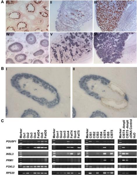
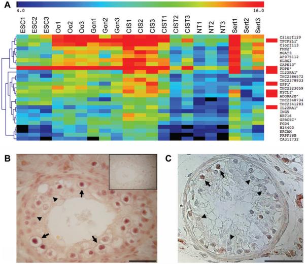
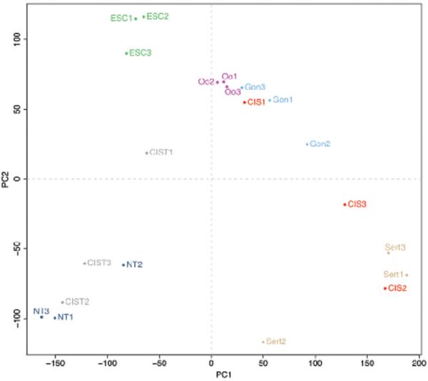
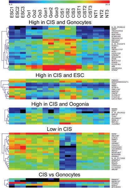
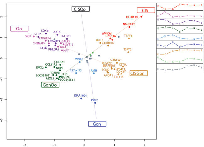
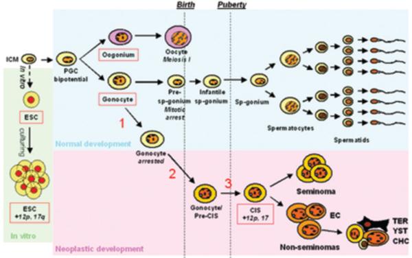
Testicular germ cell cancers in young adult men derive from a precursor lesion called carcinoma in situ (CIS) of the testis. CIS cells were suggested to arise from primordial germ cells or gonocytes. However, direct studies on purified samples of CIS cells are lacking. To overcome this problem, we performed laser microdissection of CIS cells. Highly enriched cell populations were obtained and subjected to gene expression analysis. The expression profile of CIS cells was compared with microdissected gonocytes, oogonia, and cultured embryonic stem cells with and without genomic aberrations. Three samples of each tissue type were used for the analyses. Unique expression patterns for these developmentally very related cell types revealed that CIS cells were very similar to gonocytes because only five genes distinguished these two cell types. We did not find indications that CIS was derived from a meiotic cell, and the similarity to embryonic stem cells was modest compared with gonocytes. Thus, we provide new evidence that the molecular phenotype of CIS cells is similar to that of gonocytes. Our data are in line with the idea that CIS cells may be gonocytes that survived in the postnatal testis. We speculate that disturbed development of somatic cells in the fetal testis may play a role in allowing undifferentiated cells to survive in the postnatal testes. The further development of CIS into invasive germ cell tumors may depend on signals from their postpubertal niche of somatic cells, including hormones and growth factors from Leydig and Sertoli cells.


Figures

Figure 1
Figure 1. Verification of microdissection
A: Serial sections of a testis with CIS (I and IV), fetal testis (II and V) and fetal ovary (III and VI). Top: immunohistochemical staining for AP-2γ and bottom: alkaline phophatase expression visualized by NBT-BCIP staining. B: Frozen section with CIS stained with NBT-BCIP, before (I) and after (II) laser microdissection. C: RT-PCR on representative genes. Tissues 1-3 are the same samples used in the microarray analysis, 4 and 5 are amplified RNA from microdissected and total tissues, in the left panel, CIS 4 and 5 are unamplified whole testis samples from the same patients as in the previous panel (See Table 1 for a more thorough description). Abbreviations: Oo, microdissected oogonia; FetO, fetal ovary; Gon, microdissected gonocytes; FetT, fetal testis; CIS, microdissected carcinoma in situ; CIST, testis tissue containing CIS; -Ampli, not amplified.
Figure 2
Figure 2. Verification of microarray data, re-finding CIS genes
A: 2-way SAM on CIS vs NT (delta = 0.84, FDR=0.1%) showing genes highly expressed in CIS, note that IL22RA1 is detected with 2 different oligo probes. Genes marked with red were previously described in CIS or undifferentiated testicular tumours and cell lines. The annotated genes with confirmed probe specificity are marked by asterisks. The color key at the top shows the relative expression levels from 4 to 16. A more extensive gene list (FDR<1%) can be found in Supplementary Table 2. B: Expression of MYCL1 in CIS testis. C: Expression of the THC2340734 transcript in CIS testis. Arrows indicate CIS cells and arrowheads mark Sertoli cells. Note that MYCL1 is also expressed in interstitial Leydig cells. Abbreviations: as in Fig. 1 and ESC, cultured embryonic stem cells; NT, normal testis and Sert, microdissected Sertoli cells next to CIS.
Figure 3
Figure 3. Principal component analysis
All the samples included in the microarray dataset were subjected to a principal component analysis to determine their spatial relationship to each other. Abbreviations: ESC, cultured embryonic stem cells; Oo, microdissected oogonia; Gon, microdissected gonocytes; CIS, microdissected carcinoma in situ; CIST, testis tissue containing CIS; NT, normal testis and Sert, microdissected Sertoli cells next to CIS.
Figure 4
Figure 4. Biologically interesting clusters
A 2-way SAM of interesting cell combinations. CIST was excluded in all analyses. From the top: CIS + Gonocytes vs. others (delta=1.21, FDR=0.314%), CIS + ESC vs. others (delta=1.24, FDR=0.964%), CIS + Oogonia vs. others (delta=1.33, FDR=0%), CIS vs. others (delta=3.27, FDR=0%) and CIS vs. Gonocytes (delta=3.90, FDR=0%). Annotated genes with confirmed probe specificity are marked by asterisks. The color key at the top shows the relative expression levels from 4 to 16. Abbreviations as in previous figures. Only the most differentially expressed genes are shown in Figure 4, more extensive analyses are available in supplementary Table 3.
Figure 5
Figure 5. Correspondence analysis of genes characterizing CIS, gonocytes and oogonia
Correspondance analysis for the 62 most significant genes from a three-way significance analysis of microarrays (SAM) on the classes CIS, gonocytes and oogonia. The 62 genes were clustered into nine distinct gene groups (visualized by colouring), and the corresponding mean cluster profiles are displayed as legend. The positions of the cluster names (CIS, CISOo, etc.) represent the ideal profiles for genes expressed only in the particular cell types. Δ, Gene located on the Y chromosome. Abbreviations: E, embryonic stem cells; O, oogonia; G, gonocytes; C, carcinoma in situ; T, testis tissue containing CIS; N, normal testis; S, Sertoli cells next to CIS; CISOo, CIS and oogonia; GonOo, gonocytes and oogonia and CISGon, CIS and gonocytes.
Figure 6
Figure 6. Schematic illustration of the hypothesised origin of CIS from gonocytes
Schematic illustration of the normal male germ cell development and possible transformation to CIS. ESCs are derived from the inner cell mass of a blastocyst. Prolonged culturing often leads to an accumulation of chromosomal aberrations, especially gain of material from chromosomes 12p and 17q. During early development, primordial germ cells migrate to the gonadal ridge and develop along the female (oogonia, top) or the male (gonocytes, bottom) germ cell lineage. In the male, the gonocytes become embedded in Sertoli cells, creating testicular cords. During the period from third trimester to 3 months postnatally, the gonocytes migrate to the periphery of the tubules and differentiate to pre-spermatogonia. After puberty, the spermatogonia proliferate and start spermatogenesis. CIS cells are proposed to arise when gonocytes fail to differentiate to pre-spermatogonia (1) and fail to undergo apoptosis (2). These gonocytes or pre-CIS cells lie dormant in the testis through infancy, while genomic aberrations may occur (3), and at puberty when testosterone levels increase, they start to proliferate and genomic aberrations accumulate, especially of chromosome 12 p and 17, eventually resulting in the formation of an overt tumor. Abbreviations: ICM, inner cell mass; ESC, embryonic stem cells; PGC, primordial germ cells; Sp-gonium, spermatogonium; EC, embryonal carcinoma; TER, teratoma; YST, yolk sac tumor; CHC, chorioncarcinoma.

References

    1. Rorth M, Rajpert-De Meyts E, Andersson L, Dieckmann KP, Fossa SD, Grigor KM, Hendry WF, Herr HW, Looijenga LH, Oosterhuis JW, Skakkebaek NE. Carcinoma in situ in the testis. Scand J Urol Nephrol Suppl. 2000;(205):166–86. - PubMed
    1. Skakkebaek NE, Berthelsen JG, Muller J. Carcinoma-in-situ of the undescended testis. Urol Clin North Am. 1982 Oct;9(3):377–85. - PubMed
    1. Skakkebaek NE, Rajpert-De Meyts E, Main KM. Testicular dysgenesis syndrome: an increasingly common developmental disorder with environmental aspects. Hum Reprod. 2001 May;16(5):972–8. - PubMed
    1. Sonne SB, Kristensen DM, Novotny GW, Olesen IA, Nielsen JE, Skakkebaek NE, Rajpert-De Meyts E, Leffers H. Testicular dysgenesis syndrome and the origin of carcinoma in situ testis. Int J Androl. 2008 Apr;31(2):275–87. - PubMed
    1. Holstein AF, Korner F. Light and electron microscopical analysis of cell types in human seminoma. Virchows Arch A Pathol Anat Histol. 1974;363(2):97–112. - PubMed

Publication types