. 2009 Aug;297(2):E495-504.
doi: 10.1152/ajpendo.90424.2008. Epub 2009 Jun 2.

Effects of exercise training on subcutaneous and visceral adipose tissue in normal- and high-fat diet-fed rats

Affiliations

Effects of exercise training on subcutaneous and visceral adipose tissue in normal- and high-fat diet-fed rats

Katja S C Gollisch et al. Am J Physiol Endocrinol Metab. 2009 Aug.

Abstract

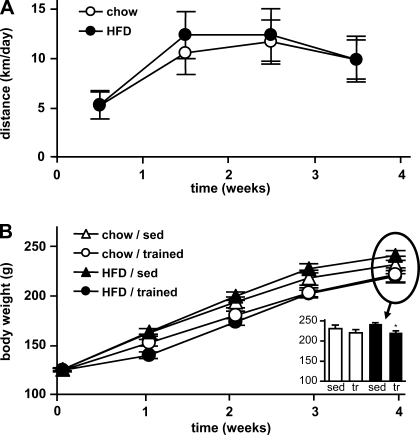
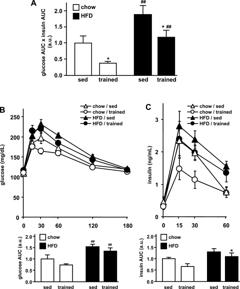
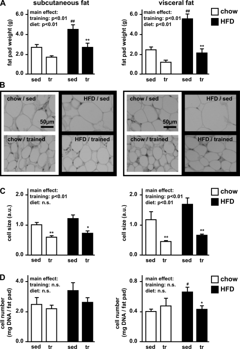
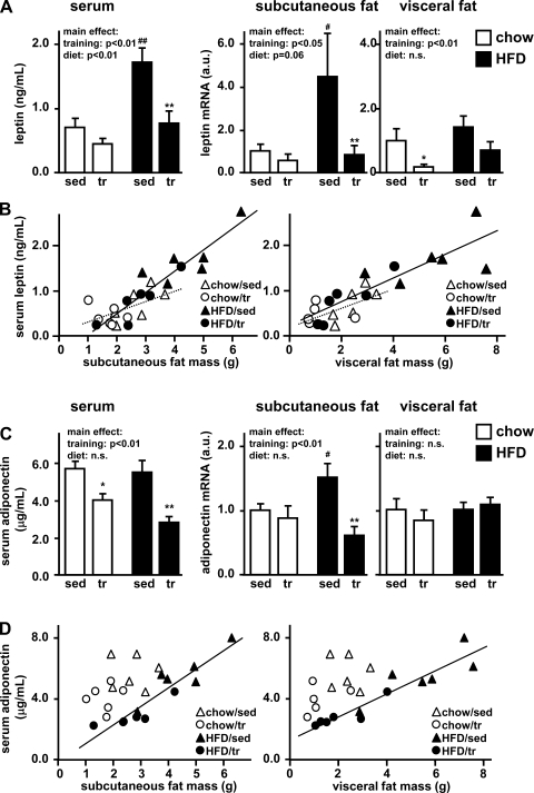
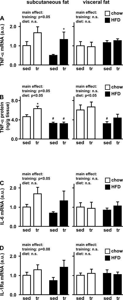
Regular physical activity improves glucose tolerance and decreases adiposity. Our aim was to investigate the effects of exercise training on subcutaneous (inguinal) and visceral (parametrial) adipose tissue in rats that were fed a chow diet (13% fat) or made insulin resistant by a high-fat diet (60% fat). Sprague-Dawley rats performed 4 wk of voluntary wheel running or were kept as sedentary controls. The training groups fed chow and the high-fat diet achieved similar running distances (8.8 +/- 1.8 and 9.3 +/- 1.9 km/day, respectively). Training improved oral glucose tolerance in chow-fed rats and prevented the glucose intolerance that occurred in sedentary rats fed the high-fat diet. In both subcutaneous and visceral adipose tissue, the high-fat diet-induced increases in fat pad weight (67% and 133%, respectively), adipocyte size (20% and 43%), and cell number (36% and 65%) were completely prevented by exercise training. Cytokine mRNA expression in visceral fat did not change with exercise training. However, in subcutaneous fat, training actually increased mRNA expression of several cytokines [IL-6: 80% (P < 0.05); TNF-alpha: 100% (P < 0.05); IL-1 receptor antagonist (IL-1Ra): 57% (P = 0.08)] with no detectable increases in serum cytokine concentrations. In summary, exercise training can overcome high-fat diet-induced impairments in glucose tolerance and increases in adipocyte size, cell number, and fat pad mass. Improved glucose tolerance was accompanied by an increase in cytokine gene expression in subcutaneous fat. This finding raises the possibility of a specific role of subcutaneous adipose tissue in adaptive responses to exercise training.


Figures

Fig. 1.
Fig. 1.
Running distance and body weights. Young female rats on regular chow or high-fat diet (HFD) were kept sedentary (sed) or performed 4 wk of voluntary wheel running (trained, tr). A: running distances in the training groups were continuously monitored over the course of 4 wk. B: body weights were determined at baseline and after 1, 2, 3, and 4 wk. Data are means ± SE. *P < 0.05 vs. sedentary.
Fig. 2.
Fig. 2.
Oral glucose tolerance. Oral glucose tolerance was assessed after 3 wk. A: as an indicator for insulin resistance, the glucose-insulin index was determined by multiplying the areas under the glucose and insulin curve (glucose AUC × insulin AUC). a.u., Arbitrary units. B: blood glucose was determined at 0, 15, 30, 60, 120, and 180 min after the glucose charge. For each group, the AUC was determined (bottom). C: serum insulin was determined at 0, 15, 30, and 60 min, and AUCs were averaged for comparison between the groups (bottom). Data are means ± SE. *P < 0.05 vs. sedentary; ##P < 0.01 vs. chow diet.
Fig. 3.
Fig. 3.
Subcutaneous and visceral fat pad weight, adipocyte size, and cell number. Adipose tissue was excised from the subcutaneous (inguinal) and visceral (parametrial) fat depots. A: adipose pad weights were determined. B: paraffin sections were stained with hematoxylin and eosin in order to visualize differences in cell size. C: adipocyte size was estimated by counting adipocytes in 10 high-power fields (HPF). Estimated relative cell size was calculated as HPF/cell number and normalized to chow/sedentary in subcutaneous adipose tissue. D: total cell number was estimated by fluorospectrometric assessment of DNA. Results are expressed as mg DNA/g fat pad. Data are as means ± SE. *P < 0.05 vs. sedentary; **P < 0.01 vs. sedentary; #P < 0.05 vs. chow diet; ##P < 0.01 vs. chow diet.
Fig. 4.
Fig. 4.
Triacylglycerol content in liver and triceps muscle. A: paraffin sections of liver were stained with hematoxylin and eosin. Triacylglycerol accumulation can be detected in the form of unstained lipid droplets in the high-fat diet groups (right). B and C: triacylglycerol concentrations in liver (B) and triceps muscle (C) were estimated by colorimetric measurements of glycerol released after ethanolic KOH hydrolysis. Data are means ± SE. **P < 0.01 vs. sedentary; ##P < 0.01 vs. chow diet.
Fig. 5.
Fig. 5.
Leptin and adiponectin. A and C: after 4 wk of exercise training and high-fat feeding, serum levels of leptin (A) and adiponectin (C) were determined by LINCO multiplex ELISAs. Adipokine gene expression was assessed in subcutaneous and visceral adipose tissue by real-time PCR. mRNA levels were normalized to the housekeeping gene β-actin and expressed relative to the sedentary/chow group. Data are means ± SE. *P < 0.05 vs. sedentary; **P < 0.01 vs. sedentary; #P < 0.05 vs. chow diet; ##P < 0.01 vs. chow diet. n.s., Not significant. B and D: correlations between adipose pad mass and leptin (B) or adiponectin (D) serum levels are shown for subcutaneous (left) and visceral (right) adipose tissue.
Fig. 6.
Fig. 6.
Macrophage markers—CD68 and monocyte chemotactic protein 1 (MCP-1). As markers for macrophage infiltration into adipose tissue, the macrophage marker gene CD68 (A) and MCP-1 (B) were assessed in subcutaneous and visceral adipose tissue by quantitative PCR. mRNA levels were normalized to the housekeeping gene β-actin and expressed relative to the respective sedentary/chow group. Data are means ± SE.
Fig. 7.
Fig. 7.
TNF-α, IL-6, and IL-1 receptor antagonist (IL-1Ra). After 4 wk of voluntary wheel running, subcutaneous and visceral adipose tissue of chow- and high fat-fed rats was studied for TNF-α (A), IL-6 (C), and IL-1Ra (D) gene expression by quantitative PCR. mRNA levels were normalized to the housekeeping gene β-actin and expressed relative to the respective sedentary/chow group. Adipose tissue TNF-α protein levels (B) were assessed by ELISA and are expressed as nanograms of protein per gram of tissue weight. Data are means ± SE. *P < 0.05 vs. sedentary; #P < 0.05 vs. chow diet.

References

    1. Atzmon G, Yang XM, Muzumdar R, Ma XH, Gabriely I, Barzilai N. Differential gene expression between visceral and subcutaneous fat depots. Horm Metab Res 34: 622–628, 2002. - PubMed
    1. Chennaoui M, Drogou C, Gomez-Merino D. Effects of physical training on IL-1beta, IL-6 and IL-1ra concentrations in various brain areas of the rat. Eur Cytokine Netw 19: 8–14, 2008. - PubMed
    1. Cortez MY, Torgan CE, Brozinick JT Jr, Ivy JL. Insulin resistance of obese Zucker rats exercise trained at two different intensities. Am J Physiol Endocrinol Metab 261: E613–E619, 1991. - PubMed
    1. Craig BW, Hammons GT, Garthwaite SM, Jarett L, Holloszy JO. Adaptation of fat cells to exercise: response of glucose uptake and oxidation to insulin. J Appl Physiol 51: 1500–1506, 1981. - PubMed
    1. Di Gregorio GB, Hensley L, Lu T, Ranganathan G, Kern PA. Lipid and carbohydrate metabolism in mice with a targeted mutation in the IL-6 gene: absence of development of age-related obesity. Am J Physiol Endocrinol Metab 287: E182–E187, 2004. - PubMed

Publication types

MeSH terms