. 2009;60(12):3337-52.
doi: 10.1093/jxb/erp167. Epub 2009 Jun 2.

A bacterial signal peptide is functional in plants and directs proteins to the secretory pathway

Affiliations

A bacterial signal peptide is functional in plants and directs proteins to the secretory pathway

Lorena Moeller et al. J Exp Bot. 2009.

Abstract

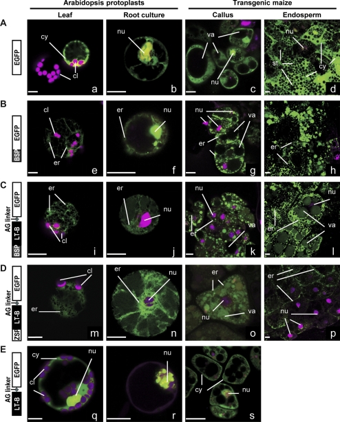
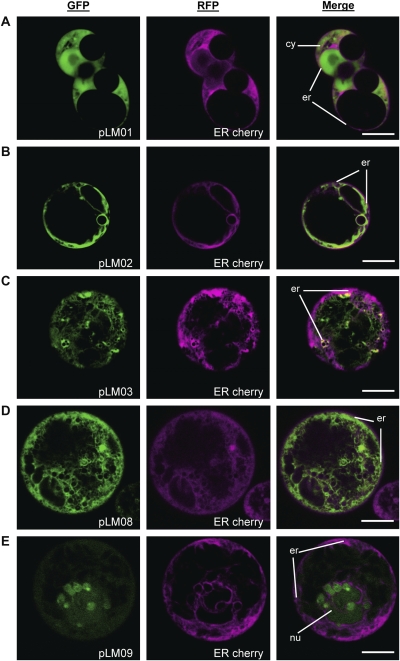
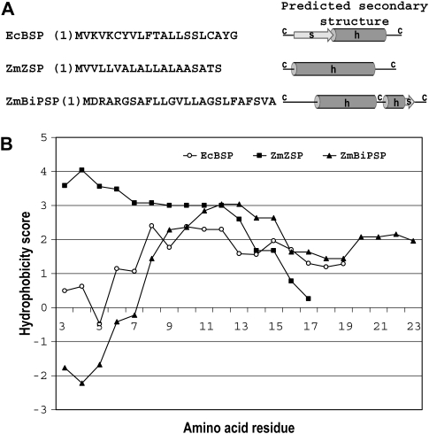
The Escherichia coli heat-labile enterotoxin B subunit (LT-B) has been used as a model antigen for the production of plant-derived high-valued proteins in maize. LT-B with its native signal peptide (BSP) has been shown to accumulate in starch granules of transgenic maize kernels. To elucidate the targeting properties of the bacterial LT-B protein and BSP in plant systems, the subcellular localization of visual marker green fluorescent protein (GFP) fused to LT-B and various combinations of signal peptides was examined in Arabidopsis protoplasts and transgenic maize. Biochemical analysis indicates that the LT-B::GFP fusion proteins can assemble and fold properly retaining both the antigenicity of LT-B and the fluorescing properties of GFP. Maize kernel fractionation revealed that transgenic lines carrying BSP result in recombinant protein association with fibre and starch fractions. Confocal microscopy analysis indicates that the fusion proteins accumulate in the endomembrane system of plant cells in a signal peptide-dependent fashion. This is the first report providing evidence of the ability of a bacterial signal peptide to target proteins to the plant secretory pathway. The results provide important insights for further understanding the heterologous protein trafficking mechanisms and for developing effective strategies in molecular farming.


Figures

Fig. 1.
Fig. 1.
Gene cassettes, plasmids, and transgenic lines for studying localization of LT-B using GFP as a reporter. Gene cassettes A–E were used in transient assays using Arabidopsis leaf and root protoplasts, and in stable transformation of maize callus and endosperm tissues. Constitutive expression in Arabidopsis protoplasts and in maize callus was driven by the double CaMV 35S promoter (P35S promoter). The maize 27 kDa γ-zein promoter (Pγzein promoter) was used to drive expression of gene cassettes A–E in maize endosperm. The number of independent transgenic events for each line is presented in parenthesis. TEV, tobacco etch virus translational enhancer leader sequence; EGFP, enhanced green fluorescent protein; Tvsp, the soybean vegetative storage protein terminator; BSP, the LT-B bacterial signal peptide; LT-B, the B subunit of E. coli heat-labile enterotoxin; ZSP, 27 kDa γ-zein signal peptide; AG linker, alanine-glycine linker; na, not available.
Fig. 2.
Fig. 2.
Gene expression analyses of LT-B, GFP, and LT-B::GFP fusions in transgenic maize kernels. (A) Bright field and fluorescence imaging of a representative self-pollinated ear of transgenic line P310 (Pγzein-BSP-LT-B::GFP) expressing GFP in the endosperm. A GFP-expressing kernel is marked by a white arrow. (B) LT-B levels as a percentage of total aqueous extractable protein (% LT-B/TAEP) in endosperm of P310 and P311 (Pγzein-ZSP-LT-B::GFP) kernels. Both transgenic maize carrying BSP- or ZSP-led LT-B::GFP fusion protein show the expression of functional LT-B. However, independent lines from both constructs have different levels of LT-B. (C) Western blot of TAEP extracts from transgenic callus (P308c, P35S-BSP-LT-B::GFP) and endosperms (P309, Pγzein-GFP, GFP control; P310-28, P310-32, two independent lines from P310; P311; and P315, Pγzein-BSP-GFP) using anti-GFP antibody. (D) Western blot of TAEP extracts from transgenic callus (P308c) and endosperms (P309, P310-32, and P315) using anti-LT-B antibody. (E) Western blot of immuno-precipitated samples using anti-LT-B antibody, probed with anti-GFP antibody. B73, non-transgenic maize line. P77, transgenic maize line expressing LT-B with its native bacterial signal peptide. The empty lane in (D) was a sample lost during loading. LT-B std, bacterial LT-B protein standard. EGFP std, commercial enhanced GFP standard. Arrowheads in (C), GFP. Dots in (C), possible cleavage peptides cross-react to GFP antibody. Asterisks in (C), (D), and (E), LT-B::GFP fusion. Open diamonds in (D), LT-B monomer. Closed diamond in (D), truncated LT-B::GFP fusion. Open circle in (D), LT-B multimer. Arrow in (E), commercial EGFP. Multiple EGFP bands in GFP standard may due to incomplete protein denaturation during boiling before loading.
Fig. 3.
Fig. 3.
Association of LT-B and GFP with starch and fibre fractions of transgenic maize kernels. Transgenic maize kernels expressing constructs presented in Fig. 1 were used for small-scale fractionation. Protein extraction from starch and fibre fractions were used for LT-B and GFP determination. (A) Functional LT-B content as a percentage of TAEP. Error bars correspond to the standard deviation of two technical replicates. (B) Anti-GFP Western blot of starch soluble (TAEP) and insoluble (pellet) phases. (C) Anti-GFP Western blot of fibre soluble (TAEP) and insoluble (pellet) phases. P309, Pγzein-GFP; P310, Pγzein-BSP-LT-B::GFP; P311, Pγzein-ZSP-LT-B::GFP; P315, Pγzein-BSP-GFP; GFP, commercial EGFP standard. Arrowheads, GFP. Open arrowhead, possible truncated form of GFP. Asterisks, LT-B::GFP fusion. Dots, possible cleavage peptides cross-react to GFP antibody. Multiple EGFP bands in GFP standard may be due to incomplete protein denaturation during boiling before loading.
Fig. 4.
Fig. 4.
Western blot analysis of total proteins from starch samples treated with Thermolysin. Starch samples were treated with Thermolysin to test the susceptibility to the protease and possible internalization of fusion proteins in the starch granules of maize. SDS-loading buffer was used to extract proteins from Thermolysin-treated starch granules, and boiled for 5 min. Samples were separated on a 12% SDS-PAGE, transferred to a 0.45 μm nitrocellulose membrane, and probed with goat anti-GFP antibodies (A), rabbit anti-waxy protein antibodies (B), or rabbit anti-27 kDa γ-zein protein antibies (C), respectively. P309, Pγzein-GFP; P310, Pγzein-BSP-LT-B::GFP; P315, Pγzein-BSP-GFP; GFP, commercial EGFP. S, starch fraction; F, fibre fraction. In (A) arrowheads, GFP; asterisks, LT-B::GFP fusion; dots, possible cleavage peptides cross-react to GFP antibody; arrows, Thermolysin-sensitive GFP band from P315 fibre fraction. Block arrow in (B), waxy protein. Open block arrows in (C), zein proteins.
Fig. 5.
Fig. 5.
Confocal images of transiently and stably transformed Arabidopsis and maize cells expressing GFP or LT-B::GFP fusion proteins. Constructs A–E are described in Fig. 1. Transiently transformed Arabidopsis leaf (a, e, i, m, q) and root (b, f, j, n, r) protoplasts using the constitutive P35S promoter constructs were imaged 24–48 h after transformation. Stably transformed maize callus (c, g, k, o, s) also used the P35S promoter constructs. Fresh immature endosperm (12–26 d after pollination) from transgenic maize seed carrying the Pγzein promoter constructs were excised and imaged (d, h, l, p). Images are presented as merged green and red channels (presented in magenta color) for all samples. Green signal in all images corresponds to GFP. Red signal in leaf protoplasts is the autofluorescence of chlorophyll in chloroplasts. Red signal in root protoplasts corresponds to the expression of a VirD2::RFP construct, a nuclear marker. Red signal in maize callus and endosperm samples is propidium iodide used as a counter stain that labels nucleic acids. Organelle labelling: chloroplasts (cl), cytosol (cy), nucleus (nu), endoplasmic reticulum (er), vacuole (va), starch (st). Bars=10 μm.
Fig. 6.
Fig. 6.
Co-localization experiments in Arabidopsis root protoplasts. Protoplasts were co-transformed using the constructs presented in Fig. 1 and an ER marker protein fused to RFP, ER cherry (Nelson et al., 2007). (A) pLM01 (GFP control). (B) pLM02 (BSP-GFP). (C) pLM03, (BSP-LT-B::GFP). (D) pLM08 (ZSP-LT-B::GFP). (E) pLM09 (LT-B::GFP). Green channel corresponds to GFP signal. Red channels (presented in magenta color) corresponds to ER-cherry signal. Merged images are also presented. Organelle labelling: cytosol (cy), nucleus (nu), endoplasmic reticulum (er). Bars=10 μm.
Fig. 7.
Fig. 7.
Amino acid sequence, predicted secondary structure, and hydropathy plots of bacterial and plant signal peptides. (A) Amino acid sequence and predicted secondary structure. (B) Hydropathy plots. EcBSP (signal peptide of the B subunit of E. coli heat-labile enterotoxin), ZmZSP (signal peptide of the maize 27 kDa γ-zein), and ZmBiPSP (signal peptide of the maize luminal binding protein). c, coil; h, helix; s, strand. Hydropathy scores were obtained based on Kyte and Doolittle (1982) using ProtScale prediction software (Gasteiger et al., 2005). Subcellular localization prediction was performed using TargetP 1.1 (Emanuelsson et al., 2000; Nielsen et al., 1997) and iPSort (Bannai et al., 2002) software.

Similar articles

Cited by

References

    1. Abramoff MD, Magelhaes PJ, Ram SJ. Image processing with ImageJ. Biophotonics International. 2004;11:36–42.
    1. Arcalis E, Marcel S, Altmann F, Kolarich D, Drakakaki G, Fischer R, Christou P, Stoger E. Unexpected deposition patterns of recombinant proteins in post-endoplasmic reticulum compartments of wheat endosperm. Plant Physiology. 2004;136:3457–3466. - PMC - PubMed
    1. Asatsuma S, Sawada C, Itoh K, Okito M, Kitajima A, Mitsui T. Involvement of α-amylase I-1 in starch degradation in rice chloroplasts. Plant and Cell Physiology. 2005;46:858–869. - PubMed
    1. Attardi G, Schatz G. Biogenesis of mitochondria. Annual Review of Cell Biology. 1988;4:289–331. - PubMed
    1. Bannai H, Tamada Y, Maruyama O, Nakai K, Miyano S. Extensive feature detection of N-terminal protein sorting signals. Bioinformatics. 2002;18:298–305. - PubMed

Publication types

MeSH terms