. 2009 Aug;37(14):4587-602.
doi: 10.1093/nar/gkp425. Epub 2009 Jun 2.

An integrative genomics approach identifies Hypoxia Inducible Factor-1 (HIF-1)-target genes that form the core response to hypoxia

Affiliations

An integrative genomics approach identifies Hypoxia Inducible Factor-1 (HIF-1)-target genes that form the core response to hypoxia

Yair Benita et al. Nucleic Acids Res. 2009 Aug.

Abstract

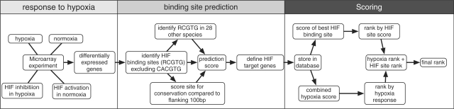
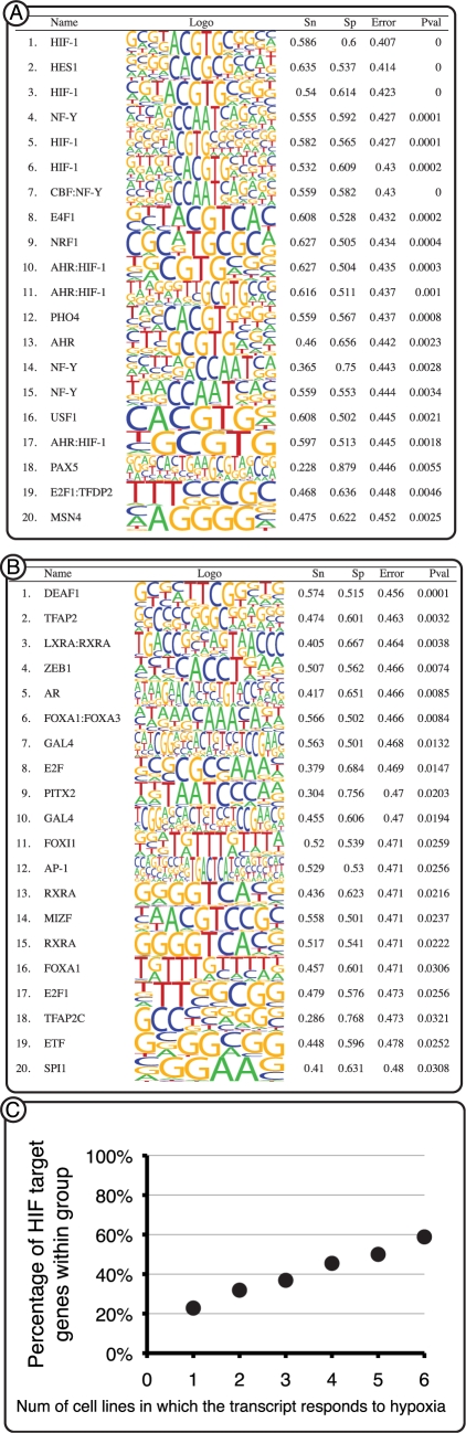
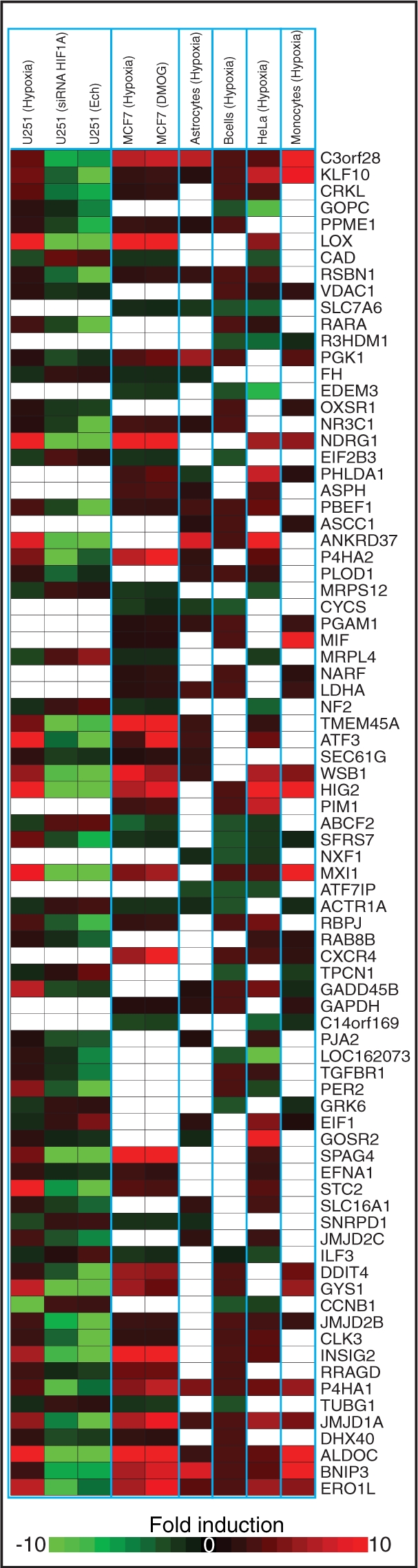
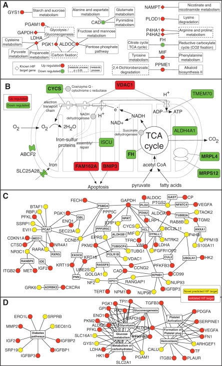
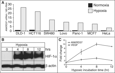
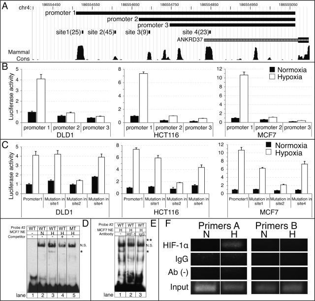
The transcription factor Hypoxia-inducible factor 1 (HIF-1) plays a central role in the transcriptional response to oxygen flux. To gain insight into the molecular pathways regulated by HIF-1, it is essential to identify the downstream-target genes. We report here a strategy to identify HIF-1-target genes based on an integrative genomic approach combining computational strategies and experimental validation. To identify HIF-1-target genes microarrays data sets were used to rank genes based on their differential response to hypoxia. The proximal promoters of these genes were then analyzed for the presence of conserved HIF-1-binding sites. Genes were scored and ranked based on their response to hypoxia and their HIF-binding site score. Using this strategy we recovered 41% of the previously confirmed HIF-1-target genes that responded to hypoxia in the microarrays and provide a catalogue of predicted HIF-1 targets. We present experimental validation for ANKRD37 as a novel HIF-1-target gene. Together these analyses demonstrate the potential to recover novel HIF-1-target genes and the discovery of mammalian-regulatory elements operative in the context of microarray data sets.


Figures

Figure 1.
Figure 1.
Prediction strategy for identifying HIF-1-target genes. Candidate genes that respond to hypoxia were first identified by microarrays. Each data set was subjected to a computational analysis in which HIF-binding sites were detected in proximal promoters. Each gene was scored for its response to hypoxia and for the best HIF-binding site. No cutoff was set for determining HIF-target genes. Finally, all genes were ranked.
Figure 2.
Figure 2.
(A and B) Binding site enrichment analysis. The program MOTIFCLASS was employed to identify binding sites significantly over-represented in the promoters of genes that respond to hypoxia in B cells. (A) Totally 770 promoters of genes that responded to hypoxia in which a detectable HIF-1-binding site was identified. HIF-1 PWMs were identified as the most enriched. (B) Totally 1456 promoters of genes that responded to hypoxia but do not contain a detectable HIF-1-binding site. No HIF-1 PWM was identified in the top 20 matrices. Sn, sensitivity; Sp, specificity; Error, classification error rate; Pval, P-value. (C) Correlation between the number of HIF-target genes and the number of cells in which they responded to hypoxia. Only 31% of the genes that responded to hypoxia in one of six cells was predicted as a HIF-target gene, compared to 71% of the genes that responded to hypoxia in six of six cell lines.
Figure 3.
Figure 3.
HIF dependent response to hypoxia across six cell types for 81 selected genes that respond to hypoxia in at least three cell types and were ranked within the top 200 HIF-target genes. The heatmap shows a fold induction compared to normoxia within each cell type. White blocks indicate the gene did not respond to hypoxia in that cell type.
Figure 4.
Figure 4.
(A) Metabolic map of HIF-target genes. The KEGG pathway database was employed to map 81 HIF-target genes defined as the core response to hypoxia to pathways. Metabolism related processes are shown here (black objects) and those over-represented with a P < 0.05 are shown with a dotted white frame. Upregulated genes are shown as red nodes while downregulated genes as green nodes. Previously identified HIF-1-target genes are indicated by a diamond shape. (B) Mitochondrial map. Genes within the top 200 predicted HIF targets that localize to the mitochondria were mapped according to their functional annotation. Upregulated genes are shown as red nodes and downregulated as green nodes. Genes indicated in bold responded to hypoxia in at least three cell types. Dotted lines from iron–sulfur protein assembly and repair illustrate a few of the iron–sulfur proteins that are key to mitochondrial function, such as NADH dehydrogenase. (C) Graphical representation of protein-protein interaction network and Reactome pathways (D) enrichment for previously validated HIF-1 targets (red) and novel predicted targets (yellow). Only proteins/pathways that were significantly enriched (P < 0.05) are shown. See Supplementary Table S6 for analysis details.
Figure 5.
Figure 5.
ANKRD37 response to hypoxia. (A) ANKRD37 mRNA levels were monitored by qPCR across seven cell lines and normalized to 18S rRNA. (B, C) ANKRD37 hypoxic induction over time during hypoxia. (B) Endogenous HIF1A protein levels determined after 0, 4, 8 and 12 h of hypoxic incubation by western blot. (C) mRNA levels of ANKRD37 and VEGFA measured by qPCR after 0, 4, 8 and 12 h of hypoxic incubation.
Figure 6.
Figure 6.
Mapping of HIF-1 site in ANKRD37 promoter. (A) Genome browser view of the four HIF-binding sites identified in the ANKRD37 promoters and the three promoter sequences that were cloned to identify the dominant HIF-1 site. The site score is shown in parenthesis for each site, higher is better. The larger box of the ANKRD37 transcript (green) indicates the coding sequence. Note that the conservation shown on the plot is computed by PhastCons which scores conservation based on the evolutionary distance and does not necessarily reflect the number of species (95). (B) DLD1, HCT116 and MCF7 cells were transiently transfected and incubated under normoxic or hypoxic conditions. Luciferase activity of the three promoters was measured and normalized to promoter 1 in normoxic conditions. All studies were performed in triplicate and data represent the mean ± SD for three different wells. (C) Luciferase activity of promoter 1 in normoxia versus hypoxia with mutations in sites 1, 2 and 4 in DLD1, HCT116 and MCF7 cells. Experimental procedure and data analysis was performed as described for panel B. (D) In vitro binding of HIF-1 protein to site 2 in the ANKRD37 promoter. EMSAs were performed using biotinylated probe corresponding to HIF-binding site 2. Nuclear extracts (NE) from MCF7 cells cultured in either normoxia (N) or hypoxia (H) were utilized. The asterisk indicates the specific shift obtained in hypoxia. 200× M excess of non-labeled probe as a specific competitor or probe with a specific mutation in the HIF-binding site2 (ACGTG > AAAAG) was utilized to confirm specificity. WT = wild-type probe. MT = mutant probe. (E) Nuclear extracts from MCF7 cells grown in hypoxia were incubated with antibody specific to HIF-1α or normal mouse IgG. The double asterisk indicates the super-shift obtained only with a HIF-1α antibody. N.S.; nonspecific band. (F) In-vivo binding of HIF-1α protein to site 2 in the ANKRD37 promoter. ChIP assays were performed using primer sets corresponding to HIF-binding site 2 (primer A) or a region upstream of site 2 (primer B). DNA–protein complexes from MCF7 cells cultured in either normoxia (N) or hypoxia (H) were immunoprecipitated with or without antibody specific to HIF-1α or normal mouse IgG. Precipitated DNA or input samples were amplified by PCR.

Similar articles

Cited by

References

    1. Wang GL, Semenza GL. Purification and characterization of hypoxia-inducible factor 1. J. Biol. Chem. 1995;270:1230–1237. - PubMed
    1. Cockman ME, Masson N, Mole DR, Jaakkola P, Chang GW, Clifford SC, Maher ER, Pugh CW, Ratcliffe PJ, Maxwell PH. Hypoxia inducible factor-alpha binding and ubiquitylation by the von Hippel-Lindau tumor suppressor protein. J. Biol. Chem. 2000;275:25733–25741. - PubMed
    1. Ohh M, Yauch RL, Lonergan KM, Whaley JM, Stemmer-Rachamimov AO, Louis DN, Gavin BJ, Kley N, Kaelin WG, Iliopoulos O. The von Hippel-Lindau tumor suppressor protein is required for proper assembly of an extracellular fibronectin matrix. Mol. Cell. 1998;1:959–968. - PubMed
    1. Kamura T, Sato S, Iwai K, Czyzyk-Krzeska M, Conaway RC, Conaway JW. Activation of HIF1alpha ubiquitination by a reconstituted von Hippel-Lindau (VHL) tumor suppressor complex. Proc. Natl Acad. Sci. USA. 2000;97:10430–10435. - PMC - PubMed
    1. Tanimoto K, Makino Y, Pereira T, Poellinger L. Mechanism of regulation of the hypoxia-inducible factor-1 alpha by the von Hippel-Lindau tumor suppressor protein. EMBO J. 2000;19:4298–4309. - PMC - PubMed

Publication types

Substances