

Fanca-/- hematopoietic stem cells demonstrate a mobilization defect which can be overcome by administration of the Rac inhibitor NSC23766
- PMID: 19491337
- PMCID: PMC2704313
- DOI: 10.3324/haematol.2008.004077
Fanca-/- hematopoietic stem cells demonstrate a mobilization defect which can be overcome by administration of the Rac inhibitor NSC23766
Abstract
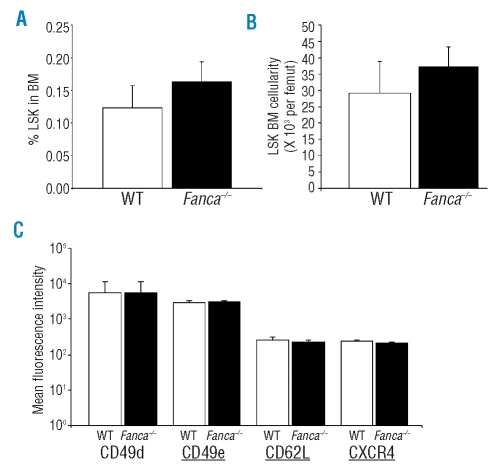
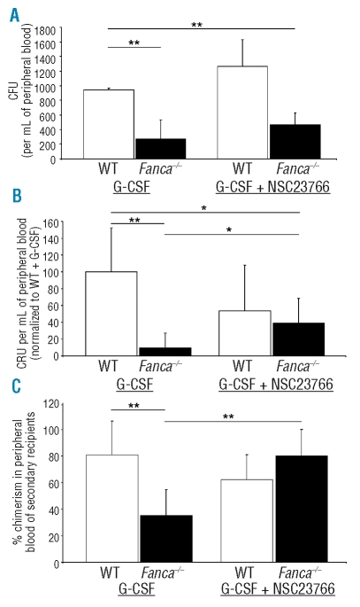
Fanconi anemia is a severe bone marrow failure syndrome resulting from inactivating mutations of Fanconi anemia pathway genes. Gene and cell therapy trials using hematopoietic stem cells and progenitors have been hampered by poor mobilization of HSC to peripheral blood in response to G-CSF. Using a murine model of Fanconi anemia (Fanca(-/-) mice), we found that the Fanca deficiency was associated with a profound defect in hematopoietic stem cells and progenitors mobilization in response to G-CSF in absence of bone marrow failure, which correlates with the findings of clinical trials in Fanconi anemia patients. This mobilization defect was overcome by co-administration of the Rac inhibitor NSC23766, suggesting that Rac signaling is implicated in the retention of Fanca(-/-) hematopoietic stem cells and progenitors in the bone marrow. In view of these data, we propose that targeting Rac signaling may enhance G-CSF-induced HSC mobilization in Fanconi anemia.
Figures
Similar articles
-
Rac GTPases differentially integrate signals regulating hematopoietic stem cell localization.Nat Med. 2005 Aug;11(8):886-91. doi: 10.1038/nm1274. Epub 2005 Jul 17. Nat Med. 2005. PMID: 16025125
-
In vitro phenotypic correction of hematopoietic progenitors from Fanconi anemia group A knockout mice.Blood. 2002 Sep 15;100(6):2032-9. Blood. 2002. PMID: 12200363
-
AMD3100 synergizes with G-CSF to mobilize repopulating stem cells in Fanconi anemia knockout mice.Exp Hematol. 2008 Sep;36(9):1084-90. doi: 10.1016/j.exphem.2008.03.016. Epub 2008 May 20. Exp Hematol. 2008. PMID: 18495331 Free PMC article.
-
Rho GTPases and regulation of hematopoietic stem cell localization.Methods Enzymol. 2008;439:365-93. doi: 10.1016/S0076-6879(07)00427-2. Methods Enzymol. 2008. PMID: 18374178 Review.
-
The role of chemokine activation of Rac GTPases in hematopoietic stem cell marrow homing, retention, and peripheral mobilization.Exp Hematol. 2006 Aug;34(8):976-85. doi: 10.1016/j.exphem.2006.03.016. Exp Hematol. 2006. PMID: 16863904 Review.
Cited by
-
Interleukin 8/KC enhances G-CSF induced hematopoietic stem/progenitor cell mobilization in Fancg deficient mice.Stem Cell Investig. 2014 Oct 31;1:19. doi: 10.3978/j.issn.2306-9759.2014.10.02. eCollection 2014. Stem Cell Investig. 2014. PMID: 27358865 Free PMC article.
-
Gata2 noncoding genetic variation as a determinant of hematopoietic stem/progenitor cell mobilization efficiency.Blood Adv. 2023 Dec 26;7(24):7564-7575. doi: 10.1182/bloodadvances.2023011003. Blood Adv. 2023. PMID: 37871305 Free PMC article.
-
Vasculopathy-associated hyperangiotensinemia mobilizes haematopoietic stem cells/progenitors through endothelial AT₂R and cytoskeletal dysregulation.Nat Commun. 2015 Jan 9;6:5914. doi: 10.1038/ncomms6914. Nat Commun. 2015. PMID: 25574809 Free PMC article.
-
The Rac GTPase effector p21-activated kinase is essential for hematopoietic stem/progenitor cell migration and engraftment.Blood. 2013 Mar 28;121(13):2474-82. doi: 10.1182/blood-2012-10-460709. Epub 2013 Jan 18. Blood. 2013. PMID: 23335370 Free PMC article.
-
Defective endomitosis during megakaryopoiesis leads to thrombocytopenia in Fanca-/- mice.Blood. 2014 Dec 4;124(24):3613-23. doi: 10.1182/blood-2014-01-551457. Epub 2014 Sep 26. Blood. 2014. PMID: 25261197 Free PMC article.
References
-
- Williams DA. Fanconi’s anemia: genomic instability leading to aplastic anemia and cancer predisposition. Hematology - European Hematol Assoc Prog. 2006;2:24–8.
-
- Garcia-Higuera I, Taniguchi T, Ganesan S, Meyn MS, Timmers C, Hejna J, et al. Interaction of the Fanconi anemia proteins and BRCA1 in a common pathway. Mol Cell. 2001;7:249–62. - PubMed
-
- Bagby GC, Alter BP. Fanconi anemia. Sem Hematol. 2006;43:147–56. - PubMed
Publication types
MeSH terms
Substances
LinkOut - more resources
Full Text Sources
Other Literature Sources
Medical
Miscellaneous
