. 2009 Jul 23;114(4):891-900.
doi: 10.1182/blood-2009-01-197178. Epub 2009 Jun 2.

Blockade of interleukin-6 signaling augments regulatory T-cell reconstitution and attenuates the severity of graft-versus-host disease

Affiliations

Blockade of interleukin-6 signaling augments regulatory T-cell reconstitution and attenuates the severity of graft-versus-host disease

Xiao Chen et al. Blood. .

Abstract

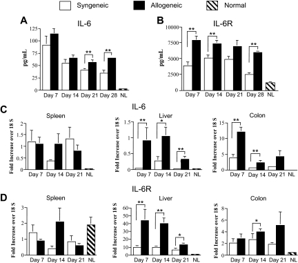
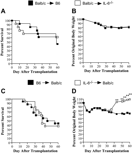
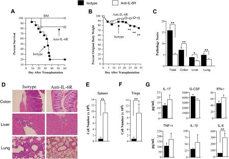
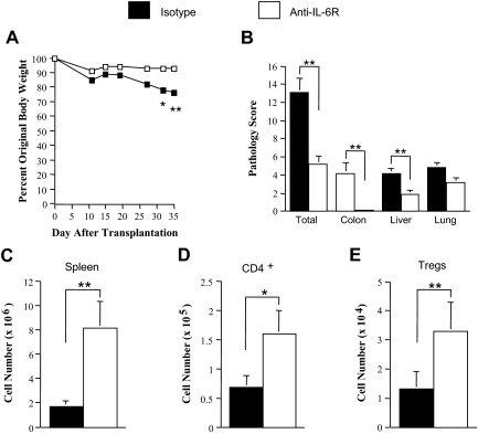
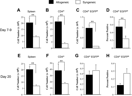
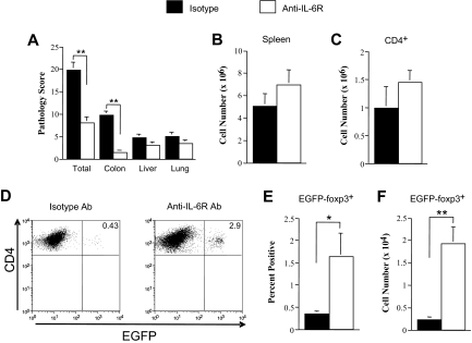
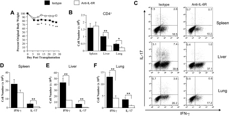
Graft-versus-host disease (GVHD) is the major complication after allogeneic bone marrow transplantation and is characterized by the overproduction of proinflammatory cytokines. In this study, we have identified interleukin-6 (IL-6) as a critical inflammatory cytokine that alters the balance between the effector and regulatory arms of the immune system and drives a proinflammatory phenotype that is a defining characteristic of GVHD. Our results demonstrate that inhibition of the IL-6 signaling pathway by way of antibody-mediated blockade of the IL-6 receptor (IL-6R) markedly reduces pathologic damage attributable to GVHD. This is accompanied by a significant increase in the absolute number of regulatory T cells (Tregs) that is due to augmentation of thymic-dependent and thymic-independent Treg production. Correspondingly, there is a significant reduction in the number of T helper 1 and T helper 17 cells in GVHD target organs, demonstrating that blockade of IL-6 signaling decreases the ratio of proinflammatory T cells to Tregs. These studies demonstrate that antibody blockade of the IL-6R serves to recalibrate the effector and regulatory arms of the immune system and represents a novel, potentially clinically translatable, strategy for the attenuation of GVHD.


Figures

Figure 1
Figure 1
IL-6 and soluble IL-6R levels are increased during GVHD. (A-B) Lethally irradiated (900 cGy) Balb/c mice received a transplant of Balb/c BM (10 × 106) and spleen cells (0.4-0.5 × 10⁶, syngeneic) or B6 BM (10 × 106) and an equivalent number of B6 spleen cells (allogeneic). Cohorts of animals (n = 5-9/group) were killed weekly and serum was analyzed for IL-6 and soluble IL-6R levels using either Bioplex or enzyme-linked immunosorbent assay as described in “Cytokine analysis.” Values for normal control mice that did not undergo transplantation (n = 5) are depicted. Data are derived from 2 independent experiments. (C-D) RNA was extracted from spleen, liver, and colon tissues obtained from recipients of syngeneic or allogeneic marrow grafts (n = 7-8 mice per tissue) at the indicated time points, and gene expression of IL-6 (C) and total IL-6R (D) was analyzed by real-time q-PCR as described in “Real-time q-PCR.” Values for normal control mice that did not undergo transplantation (n = 5-6 mice/tissue) are depicted. Data are derived from 2 independent experiments and are presented as the mean ± SEM. (Statistics: *P ≤ .05, **P < .01.)
Figure 2
Figure 2
Absence of either recipient or donor-derived IL-6 is insufficient to protect mice from lethal GVHD. (A-B) Lethally irradiated (1000 cGy) B6 (n = 13) or IL-6−/− (n = 13) animals received a transplant of 10 × 106 BM and 15 × 106 spleen cells from Balb/c mice. Overall survival (A) and the percentage of original body weight over time (B) are depicted. Data are cumulative results from 4 independent experiments. (C-D) Lethally irradiated (900 cGy) Balb/c mice received a transplant of 10 × 106 BM and 0.5 to 0.7 × 10⁶ spleen cells from either B6 (n = 16) or IL-6−/− (n = 16) animals. Overall survival (C) and the percentage of original body weight over time (D) are depicted. Data are cumulative results from 4 independent experiments. (Statistics: **P < .01.)
Figure 3
Figure 3
Antibody blockade of the IL-6R significantly attenuates the severity of GVHD. (A) Lethally irradiated (900 cGy) Balb/c mice received a transplant of T cell–depleted (TCD) B6 BM alone (10 × 106; ○, n = 9) or together with B6 spleen cells adjusted to yield a T-cell dose of 0.7 × 10⁶ αβ T cells. Cohorts of mice that received adjunctive spleen cells were then treated with either rat IgG isotype control (n = 15) or anti–IL-6R (MR-16-1) antibody (n = 12) once weekly for 4 weeks beginning on the day of transplantation. Overall survival is depicted. Data are cumulative results from 3 independent experiments. (B) Lethally irradiated Balb/c mice received a transplant of TCD Foxp3EGFP BM (10 × 106) and 0.4 to 0.5 × 10⁶ Foxp3EGFP spleen cells. Cohorts of mice were then treated with either rat IgG isotype control (n = 12) or anti–IL-6R (MR-16-1) antibody (n = 12) once weekly for 4 weeks beginning on the day of transplantation. Mice from both groups were killed 34 to 37 days after BMT. (B) The percentage of original body weight over time in mice from both groups is depicted. (C) Pathological damage in the colon, liver, and lung using a semiquantitative scoring system as detailed in “Histologic analysis.” (D) Histology of colon, liver, and lung from representative recipients treated with either isotype or anti–IL-6R antibody. In isotype control animals, colon shows extensive inflammation in the lamina propria, goblet cell depletion, and crypt cell destruction; liver reveals portal triad inflammation with mononuclear cells and endothelialitis; and lung demonstrates perivascular and peribronchial cuffing with mononuclear cells. In anti–IL-6R antibody–treated mice, colon has normal-appearing mucosa with no attendant inflammation, liver has reduced portal triad inflammation, and lung demonstrates a similar reduction in perivascular and peribronchial cuffing. (E) Total spleen cellularity and (F) absolute number of splenic Tregs (CD4+ EGFP+) are shown. (G) Lethally irradiated (900 cGy) Balb/c mice received a transplant of TCD B6 BM plus 0.4 × 10⁶ B6 spleen cells. Cohorts of animals that underwent transplantation were then treated with either isotype control (n = 12) or anti–IL-6R antibody (n = 12) on the day of transplantation. Mice were bled 6 days after transplantation and serum was assayed for proinflammatory cytokines. Data are presented as the mean (± SEM) and are the cumulative results from 3 independent experiments. (Statistics: *P ≤ .05, **P < .01.)
Figure 4
Figure 4
Attenuation of GVHD by blockade of IL-6 signaling does not require an intact thymus. Lethally irradiated (900 cGy) thymectomized Balb/c mice received a transplant of Foxp3EGFP BM (10 × 106) and 0.4 × 10⁶ Foxp3EGFP spleen cells. Cohorts of mice were then treated with either rat IgG isotype control (n = 12) or anti–IL-6R antibody (n = 12) once weekly for 4 weeks beginning on the day of transplantation. Animals were then killed 46 to 48 days after transplantation. (A) The percentage of original body weight of mice over time from both groups is depicted. (B) Pathological damage in the colon, liver, and lung using a semiquantitative scoring system as detailed in “Histologic analysis.” (C) Total spleen cellularity, (D) absolute number of splenic CD4+ T cells, and (E) absolute number of splenic Tregs are depicted. Data are presented as the mean (± SEM) and are the cumulative results from 3 independent experiments. (Statistics: *P ≤ .05, **P < .01.)
Figure 5
Figure 5
In vivo conversion of CD4+ foxp3 T cells to CD4+ foxp3+ T cells is negligible during acute GVHD. Lethally irradiated bm12 (1000 cGy; n = 8-11/group) or B6 (1000 cGy; n = 8-12/group) mice received a transplant of B6 Rag-1 BM (5 × 10⁶) and sorted CD4+ EGFP-foxp3 T cells (0.6 × 10⁶). Cohorts of mice were killed on either days 7 to 9 or day 20 after transplantation. Spleen cellularity (A,E), absolute number of splenic CD4+ T cells (B,F), percentage of CD4+ EGFP+ T cells in the spleen (C,G), and absolute number of CD4+ EGFP+ T cells in the spleen (D,H) are depicted. Data are presented as the mean (± SEM) and are the cumulative results from 2 to 3 independent experiments. (Statistics: **P < .01.)
Figure 6
Figure 6
Antibody blockade of the IL-6R augments conversion of CD4+ foxp3 to CD4+ foxp3+ Tregs. Lethally irradiated (900 cGy) Balb/c mice received a transplant of B6 Rag-1 BM (5 × 10⁶) and sorted CD4+ EGFP-foxp3 T cells (0.2 × 10⁶). Cohorts of mice were then administered rat IgG isotype control (n = 9) or anti–IL-6R antibody (n = 12) once weekly for 4 weeks as described in “Methods.” Mice in both groups were killed 26 to 36 days after transplantation. (A) Pathological damage in the colon, liver, and lung using a semiquantitative scoring system as detailed in “Histologic analysis.” (B) Total spleen cellularity and (C) absolute number of splenic CD4+ T cells are depicted. (D) Representative dot plot showing percentage of EGFP-foxp3+ iTregs in the gated CD4+ T-cell population from transplant recipients treated with either isotype control or anti–IL-6R antibody. (E) Percentage and (F) absolute number of iTregs in the spleen of animals administered control or anti–IL-6R antibody. Data are presented as the mean (± SEM) and are the cumulative results from 3 independent experiments. (Statistics: *P ≤ .05, **P < .01.)
Figure 7
Figure 7
Blockade of IL-6 signaling results in a significant reduction in proinflammatory TH1 and TH17 cells. Lethally irradiated (900 cGy) Balb/c mice received a transplant of B6 BM (5 × 10⁶) and purified B6 CD4+ T cells (0.3 × 10⁶). Cohorts of mice were then administered rat IgG isotype control (n = 8) or anti–IL-6R antibody (n = 8) for 4 weekly injections as described in “Reagents.” Mice in both groups were killed 28 to 29 days after transplantation. (A) The percentage of original body weight of mice over time from both groups is depicted. (B) Absolute number of CD4+ T cells in the spleen, liver, and lung of animals treated with either isotype control or anti–IL-6R antibody. (C) Representative dot plot depicting the percentage of IL-17– and/or IFN-γ–secreting cells within the gated CD4+ T-cell population. (D-F) Absolute number of CD4+ IFN-γ+ or CD4+ IL-17+ T cells present in the spleen, liver, or lung of animals treated with either isotype control or anti–IL-6R antibody. Data are presented as the mean (± SEM) and are the cumulative results from 2 independent experiments. (Statistics: *P ≤ .05, **P < .01.)

References

    1. Hill GR, Crawford JM, Cooke KR, Brinson YS, Pan L, Ferrara JL. Total body irradiation and acute graft versus host disease: the role of gastrointestinal damage and inflammatory cytokines. Blood. 1997;90:3204–3213. - PubMed
    1. Xun CQ, Thompson JS, Jennings CD, Brown SA, Widmer MB. Effect of total body irradiation, busulfan-cyclophosphamide, or cyclophosphamide conditioning on inflammatory cytokine release and development of acute and chronic graft versus host disease in H-2 incompatible transplanted SCID mice. Blood. 1994;83:2360–2367. - PubMed
    1. Ferrara JL, Reddy P. Pathophysiology of graft versus shot disease. Semin Hematol. 2006;43:3–10. - PubMed
    1. Teshima T, Ordemann R, Reddy P, et al. Acute graft versus host disease does not require alloantigen expression on host epithelium. Nat Med. 2002;8:575–581. - PubMed
    1. Mowat AC. Antibodies to IFN-γ prevent immunologically mediated intestinal damage in murine graft versus host disease. Immunology. 1989;68:18–23. - PMC - PubMed

Publication types

MeSH terms