. 2009 Aug 7;284(32):21647-58.
doi: 10.1074/jbc.M109.016923. Epub 2009 Jun 2.

Monoclonal antibodies recognize distinct conformational epitopes formed by polyglutamine in a mutant huntingtin fragment

Affiliations

Monoclonal antibodies recognize distinct conformational epitopes formed by polyglutamine in a mutant huntingtin fragment

Justin Legleiter et al. J Biol Chem. .

Abstract

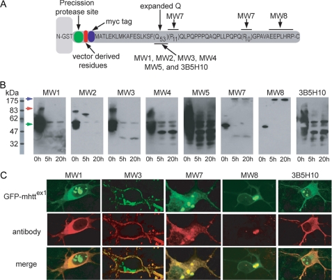
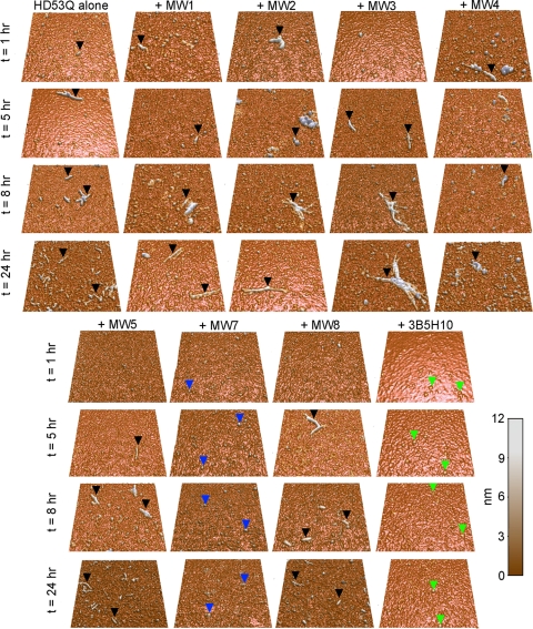
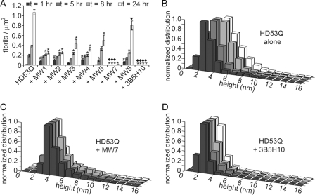
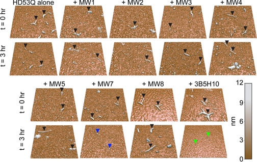
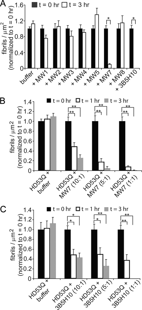
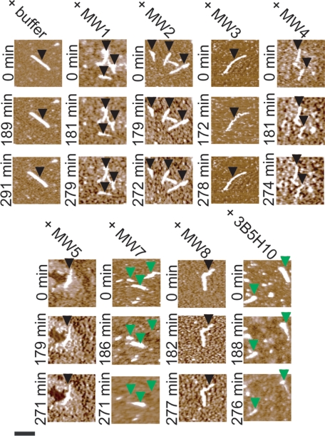
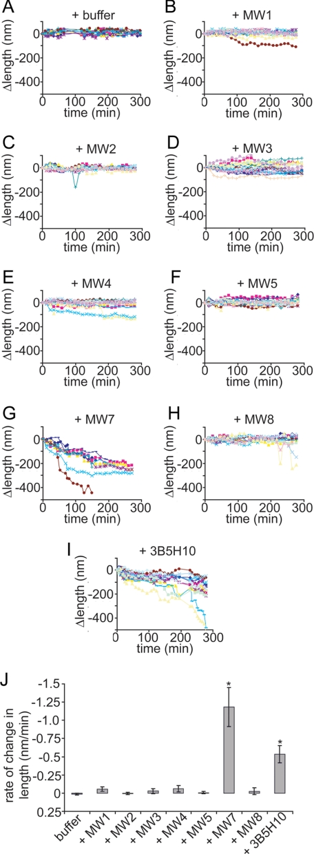
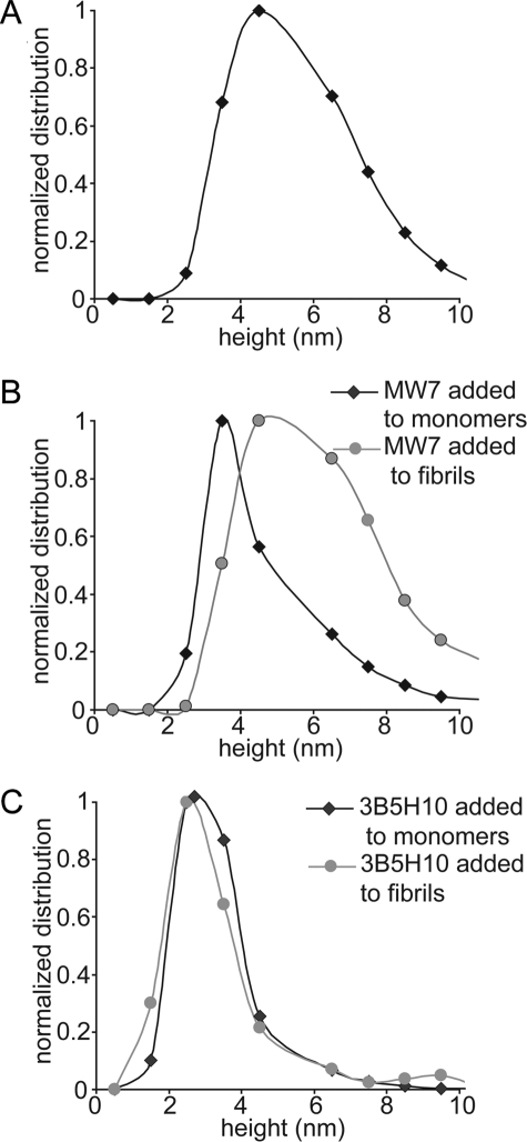
Huntington disease (HD) is a neurodegenerative disorder caused by an expansion of a polyglutamine (polyQ) domain in the N-terminal region of huntingtin (htt). PolyQ expansion above 35-40 results in disease associated with htt aggregation into inclusion bodies. It has been hypothesized that expanded polyQ domains adopt multiple potentially toxic conformations that belong to different aggregation pathways. Here, we used atomic force microscopy to analyze the effect of a panel of anti-htt antibodies (MW1-MW5, MW7, MW8, and 3B5H10) on aggregate formation and the stability of a mutant htt-exon1 fragment. Two antibodies, MW7 (polyproline-specific) and 3B5H10 (polyQ-specific), completely inhibited fibril formation and disaggregated preformed fibrils, whereas other polyQ-specific antibodies had widely varying effects on aggregation. These results suggest that expanded polyQ domains adopt multiple conformations in solution that can be readily distinguished by monoclonal antibodies, which has important implications for understanding the structural basis for polyQ toxicity and the development of intrabody-based therapeutics for HD.


Figures

FIGURE 1.
FIGURE 1.
Anti-htt antibodies recognize a variety species of HD53Q in vitro and in situ. A, a schematic representation of the GST-htt exon 1 fusion protein with 53Q (HD53Q) shows a PreScission protease site located between GST and the htt fragment (not drawn to scale) and the locations of epitopes for the antibodies that were used in this study. B, Western blots of HD53Q after incubation with protease for varying times, probed with antibodies as labeled. The location of bands representing intact GST-HD53Q fusion protein at ∼53 kDa is indicated by a green arrow. A band that migrated at an apparent molecular mass of a dimer of the fusion protein (∼106 kDa) is indicated by a red arrow. A blue arrow indicated the location of the wells of the gel where larger HD53Q aggregates are observed. C, primary cultures of rat striatal neurons expressing a GFP-labeled mutant htt-exon1 fragment with 97Q were analyzed by immunocytochemistry with antibodies as labeled.
FIGURE 2.
FIGURE 2.
Anti-htt antibodies modulate htt aggregation differentially. Representative 2 μm × 2 μm AFM images of 20 μm HD53Q incubated in the absence or presence of antibodies as labeled for 1, 5, 8, and 24 h after cleavage of the GST moiety. The ratio of antigen binding sites to HD53Q was 1:1. For HD53Q alone and with MW1-MW5 or MW8, fibrillar structures (black arrows) appeared after 1–5 h of incubation. The number of fibrils increased at 8 and 24 h. However, it appeared that there were more fibrils for HD53Q alone. For incubations of HD53Q with MW7 or 3B5H10, no fibrillar structures appeared throughout the 24-h experiment. In incubations with MW7, globular aggregates (blue arrows) around ∼3.5 nm tall were the dominant species observed at all time points. For incubation with 3B5H10, smaller globular species (green arrows) ∼2.5 nm tall were present at all time points. Shown are representative AFM images. Quantification of the number of fibrils per μm2 in these experiments is shown in Fig. 3.
FIGURE 3.
FIGURE 3.
Quantification over time of HD53Q aggregates in the absence and presence of anti-htt antibodies. A, the number of fibrils/μm2 was calculated from AFM images of HD53Q incubated in the absence and presence of anti-htt antibodies analyzed at 1, 5, 8, and 24 h of incubation. Compared with control experiments of HD53Q alone, all of the antibodies significantly reduced the number of fibrils formed at later time points. However, there was a significant increase in the number of fibrils formed after 1 h for incubations with MW1, MW2, MW4, and MW8. MW7 and 3B5H10 completely inhibited the formation of fibrils over the time course of the experiments. #, a significant increase (p < 0.05) in the number of fibrils/μm2 in comparison to HD53Q alone at the same time point (Student's t test). * and ▼ denote significant decreases (* = p < 0.01, ▼ = p < 0.05) in the number of fibrils/μm2 in comparison to HD53Q alone at the same time point (Student's t test). ♦ indicates that no fibrils were observed. The experiment was replicated six times, and the error bars represent standard error. B–D, height histograms for globular structures observed in HD53Q alone (B) and with MW7 (C) or 3B5H10 (D) as a function of time. Whereas the height of HD53Q oligomers gradually increased over time, both MW7 and 3B5H10 stabilized distinct globular structures that likely represent complexes of HD53Q and antibody. The legend applies to all panels in the figures.
FIGURE 4.
FIGURE 4.
Ex situ AFM analysis indicates that the anti-htt antibodies MW7 and 3B5H10 disassemble htt aggregates. Samples of HD53Q were incubated for 6–8 h after removal of the GST moiety to form a large population of fibrils. Then, buffer (as control), MW1-MW5, MW7, MW8, or 3B5H10 was added. The ratio of antigen binding sites to HD53Q was 1:1. The solutions were sampled directly after the addition of buffer/antibodies (t = 0 h) and deposited on mica for AFM imaging. Fibrils (black arrows) were present in all samples at this time. The solutions were incubated for 3 h after the addition of buffer or antibodies and re-sampled. Fibrils (black arrows) were still present in samples that had been treated with buffer, MW1-MW5 or MW8. However, fibrils were no longer detected in samples treated with MW7 or 3B5H10. Treatment with MW7 resulted in a large population of globular species (blue arrows) that varied greatly in size with the majority of species ranging in height from 4 to 8 nm. Treatment with 3B5H10 resulted in globular species (green arrows) that were only ∼2.5 nm tall. Shown are representative 2 μm × 2 μm AFM images. Quantification of the number of fibrils per μm2 in these experiments is shown in Fig. 5.
FIGURE 5.
FIGURE 5.
Quantification of the number of fibrils/μm2 for pre-aggregated HD53Q treated with buffer or anti-htt antibodies. A, the number of fibrils/μm2 was calculated from AFM images of incubations of fibrillar preparations of HD53Q taken immediately after (t = 0 h) and 3 h after the addition of buffer, MW1-MW5, MW7, MW8, or 3B5H10. The ratio of antigen binding sites to HD53Q was 1:1. For comparison, all bars are normalized to the number of fibrils/μm2 at t = 0 h for that sample. With the addition of buffer (control), MW1-MW5, or MW8, there was no change in the number of fibrils present after 3 h. With MW7 and 3B5H10, the number of fibrils was significantly reduced, indicating that these antibodies were able to disassemble preformed fibrils. *, p < 0.001 (Student's t test). Error bars represent standard error. B and C, the dose dependence of fibril disaggregation was studied by quantitative analysis of AFM images of fibrillar preps of HD53Q taken immediately after (t = 0 h), 1 h, and 3 h after the addition of B, MW7 or C, 3B5H10. The ratio of antigen binding sites to HD53Q was 10:1, 5:1, and 1:1. For comparison, all bars are normalized to the number of fibrils/μm2 at t = 0 h for that sample. The disaggregation of fibrils by MW7 (B) and 3B5H10 (C) appeared to be dose-dependent. *, p < 0.01; **, p < 0.001 (Student's t test).
FIGURE 6.
FIGURE 6.
Monitoring disassembly of single htt aggregates incubated with MW7 or 3B5H10 by in situ AFM. Samples of HD53Q were incubated for 6–8 h after removal of the GST moiety to form a large population of fibrils. These fibrils were deposited on mica and imaged using in situ AFM, which allows for the tracking of the fate of individual fibrils as a function of time. These fibrils were imaged in the absence or presence of anti-htt antibodies. Fibrils appeared to be stable after treatment with buffer, MW1-MW5, or MW8 (location of stable fibrils indicated by black arrows). However, treatment with MW7 or 3B5H10 caused fibrils to disaggregate and/or shorten in length (location of disaggregating fibrils indicated by green arrows). Scale bar represents 500 nm and is applicable to all images. See also supplemental movies S1 and S2.
FIGURE 7.
FIGURE 7.
Quantification of change in length and rate of change of fibrils treated with anti-htt antibodies. A–I, the change in length (Δlength) of individual fibrils imaged in the absence and presence of anti-htt antibodies was tracked as a function of time as measured by in situ AFM. Fibril length appeared stable with the addition of buffer (A), MW1-MW5 (B–F), or MW8 (H). The length of individual fibrils steadily decreased after treatment with MW7 (G) or 3B5H10 (I). J, the average rate of change of fibril length for fibrils treated with buffer (as control), MW1-MW5, MW7, MW8, or 3B5H10 was calculated, showing that only MW7 and 3B5H10 caused a significant change in fibril length (*, p < 0.01 with a Student's t test).
FIGURE 8.
FIGURE 8.
Size analysis of aggregate observed with MW7 or 3B5H10 added to monomeric or fibrillar HD53Q. A, HD53Q oligomers (HD53Q incubated alone) after 5 h of incubation were predominantly 4–5 nm in height with several as tall as 6–8 nm. B, when MW7 was incubated (added at t = 0 h) with monomeric HD53Q (black diamonds), the height of globular aggregates formed after 5 h of co-incubation were predominantly 3–4 nm tall, although there was a large portion of taller globular aggregates (shoulder on the right of the histogram). In contrast, when MW7 was incubated with pre-aggregated fibrillar HD53Q (gray circles), globular aggregates (conditions where fibrils disaggregated) observed when imaged 3 h after addition of MW7 were much taller (4–5 nm) in comparison to those formed by adding MW7 to monomeric HD53Q, with a larger portion of aggregates being 5–10 nm tall. C, when 3B5H10 was incubated (added at t = 0 h) with monomeric HD53Q (black diamonds), the majority of globular aggregates observed after 5 h co-incubation were 2–3 nm in height. Similarly, when 3B5H10 was incubated with pre-aggregated fibrillar HD53Q (gray circles), globular species (conditions where fibrils disaggregated) observed 3 h after the addition of 3B5H10 again were predominantly 2–3 nm tall.

References

    1. Vonsattel J. P., Myers R. H., Stevens T. J., Ferrante R. J., Bird E. D., Richardson E. P., Jr. (1985) J. Neuropathol. Exp. Neurol. 44, 559–577 - PubMed
    1. Buxbaum J. N. (2003) Trends Biochem. Sci. 28, 585–592 - PubMed
    1. Chiti F., Dobson C. M. (2006) Annu. Rev. Biochem. 75, 333–366 - PubMed
    1. Ross C. A., Poirier M. A. (2004) Nat. Med. 10, S10–S17 - PubMed
    1. Tobin A. J., Signer E. R. (2000) Trends Cell Biol. 10, 531–536 - PubMed

Publication types