Review
. 2009 Jul;10(7):495-506.
doi: 10.1038/nrn2636. Epub 2009 Jun 3.

The diverse functional roles and regulation of neuronal gap junctions in the retina

Affiliations
Review

The diverse functional roles and regulation of neuronal gap junctions in the retina

Stewart A Bloomfield et al. Nat Rev Neurosci. 2009 Jul.

Abstract

Electrical synaptic transmission through gap junctions underlies direct and rapid neuronal communication in the CNS. The diversity of functional roles that electrical synapses have is perhaps best exemplified in the vertebrate retina, in which gap junctions are formed by each of the five major neuron types. These junctions are dynamically regulated by ambient illumination and by circadian rhythms acting through light-activated neuromodulators such as dopamine and nitric oxide, which in turn activate intracellular signalling pathways in the retina.The networks formed by electrically coupled neurons are plastic and reconfigurable, and those in the retina are positioned to play key and diverse parts in the transmission and processing of visual information at every retinal level.


Figures

Figure 1
Figure 1. Structure and molecular organization of gap junctions
a | Gap junctions are formed between the opposing membranes of neighbouring cells. Hemichannels on each side dock to one another to form conductive channels between the two cells. An extended field of these channels forms a gap junctional plaque. Each hemichannel, or connexon, is comprised of 6 connexin protein subunits that are oriented perpendicular to the cells' membranes to form a central pore. This central pore serves as a conduit for ions and low-molecular-mass molecules of up to 1,000 Da. Connexons can contain only one type of connexin subunit (homomeric connexons) or a mixture of different connexins (heteromeric connexons). Gap junctional channels can consist of two of the same connexon (homotypic channels) or of connexons with different subunit compositions (heterotypic channels). b | Connexin subunits are proteins that have four transmembrane domains, two extracellular loops (E1 and E2) and one intracellular loop, as well as carboxyl and amino termini in the cytoplasm. Although the four transmembrane domains (M1–M4) share a conservative sequence that is important for docking in the cellular membrane, their cytoplasmic domains vary in length and amino acid sequence. Regulation of the three-dimensional connexin structure, which underlies the opening and closing of gap junction channels, is mediated at the cytoplasmic regions.
Figure 2
Figure 2. Neuromodulators affect gap junction conductances through intracellular pathways
A summary of the intracellular pathways by which dopamine (DA) and nitric oxide (NO) are thought to affect the conductance of retinal gap junctions. DA release from dopaminergic amacrine cells is increased by light. For some retinal neurons DA binds to D1 receptors (left panel), activating adenylate cyclase (ACy) and increasing the concentration of cyclic AMP. This in turn activates cAMP-dependent protein kinase (PKA), the phosphorylation of connexins and a reduction in the conductance (g) of gap junctions. (However, it has recently been suggested that in AII amacrine cells PKA activates a phosphatase (Ph) that dephosphorylates connexins and thereby causes reduced gap junction conductance) This D1 receptor mechanism occurs at gap junctions between horizontal cells (HC–HC),, between AII amacrine cells (AII–AII),,, between other amacrine cell subtypes (AC–AC) and at the amacrine cell hemichannel of amacrine cell–ganglion cell gap junctions (AC–GC). DA also binds to D2/4 receptors (middle panel), which reduces the activity of adenylate cyclase, resulting in a reduction of cAMP levels. This reduces the activity of PKA, resulting in increased gap junction conductance. This mechanism occurs at gap junctions between rods and cones (R–C), between ganglion cells (GC–GC) and at the ganglion cell hemichannel of ganglion cell–amacrine cell gap junctions (GC–AC). Light also increases the release of NO from NADPH/nitric oxide synthase (NOS)-positive amacrine cells (right panel). NO diffuses into retinal neurons and activates guanylate cyclase (GCy), resulting in an increase in cGMP levels, activation of a cGMP-dependent protein kinase (PKG), phosphorylation of connexins and reduced gap junction conductance. This mechanism occurs at gap junctions between horizontal cells (HC–HC),, and between AII amacrine cells and ON cone bipolar cells (AII–CB).
Figure 3
Figure 3. Gap junctions expressed by retinal neurons
This schematic shows seven examples of electrical coupling, the different functions of which are detailed in the main text. The coloured ovals represent gap junction hemichannels. The solid and dotted arrows represent excitatory and inhibitory chemical synapses, respectively. a | Both hemichannels of the gap junctions that couple neighbouring cones (C) express CX36 (Refs 48,49). b | In rod (R)–cone gap junctions, only the hemichannel on the cone side contains CX36; the connexin on the rod side remains unknown,,,. c | The type of connexin in rod–rod gap junctions is also unknown. d | Horizontal cell (HC) dendrites are extensively coupled. In mammals, axonless horizontal cells express CX50 (Ref. 166) whereas axon-bearing horizontal cells express CX57 (Refs 86,87). e,f | AII amacrine cells (AII) form two types of gap junction. Gap junctions between AII cells seem to be homotypic and comprised of homomeric hemichannels containing CX36 (e),,. By contrast, gap junctions between AII amacrine cells and ON cone bipolar cells (CB) can be homotypic or heterotypic, with the AII cell hemichannels containing CX36 and the cone bipolar cell hemichannel containing either CX36 or CX45 (Refs 70,121–125). g | Ganglion cells (GC) are extensively coupled to each other and/or to neighbouring amacrine cells (AC). To date, ganglion cell gap junctions have been reported to contain CX36 or CX45 (Refs 167–169). GCL; ganglion cell layer; INL, inner nuclear layer; IPL, inner plexiform layer; ONL, outer nuclear layer; OPL, outer plexiform layer; RB, rod bipolar cell.
Figure 4
Figure 4. The three rod pathways in the mammalian retina
a | The primary rod pathway involves electrical synapses between AII amacrine cells (AII), and between AII cells and cone bipolar cells (CB). In this pathway, signals are transmitted from rods (R) to rod bipolar cells (RB) and subsequently to AII cells. AII cells make sign-conserving electrical synapses with ON cone bipolar cells and sign-inverting inhibitory chemical synapses with OFF cone bipolar cells. In turn, the ON and OFF cone bipolar cells make excitatory chemical synapses with ON and OFF ganglion cells (GC), respectively. b | The secondary rod pathway involves electrical synapses between rod and cone photoreceptors (C). In this pathway, rod signals are transmitted directly from rods to cone photoreceptors through interconnecting gap junctions. The rod signals are then relayed to ON and OFF cone bipolar cells, which carry the signals to ganglion cells in the inner retina. c | The tertiary rod pathway involves electrical synapses between rods only. In this pathway, rods make direct chemical synapses with a subset of OFF bipolar cells (BC), which transmit the signals to some OFF ganglion cells. This pathway does not seem to have a counterpart in the ON circuitry. The solid and dotted arrows represent excitatory and inhibitory chemical synapses, respectively. Figure is modified, with permission, from Ref. © (2006) Society for Neuroscience.
Figure 5
Figure 5. Electrical coupling between AII amacrine cells is regulated by background light conditions
The extent of coupling between AII amacrine cells under three different background light conditions, mimicking starlight, twilight and daylight. Each group of red symbols provides the average extent of Neurobiotin tracer coupling of rabbit AII amacrine cells. Under dim starlight conditions, the conductance of the gap junctions connecting neighbouring AII amacrine cells is relatively low, and so tracer movement is limited to only a few cells. As the ambient background light increases to twilight conditions, the conductance of the gap junctions increases and so the electrical syncytium of the coupled cells enlarges dramatically. Under bright daylight conditions, the conductance of the gap junctions is once again reduced and electrical communication is limited to a small group of cells. This triphasic modulation of AII cell coupling to light is similar to that seen for horizontal cells and ensures that the fidelity of the signals carried by the primary rod pathway is maintained under different scotopic conditions. Under bright background conditions, the limited coupling between AII amacrine cells limits lateral interactions that would blur the image, thereby maintaining the high acuity that is essential for daylight vision. Figure is modified, with permission, from Ref. © (2004) Elsevier.
Figure 6
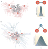
Figure 6. Ganglion cell gap junctions underlie two patterns of concerted spike activity
a | Direct electrical coupling between neighbouring ganglion cells (GC) (left panel, red and brown) results in concerted activity. Paired recordings from coupled ganglion cell neighbours generate a cross-correlogram function with a distinct pattern consisting of two peaks with short latencies of approximately 1–3 ms (right panel). These two peaks reflect reciprocal interactions in which a spike in one cell gives rise to a spike in a coupled neighbour. b | Indirect electrical coupling between ganglion cells (red and brown) through gap junctions with a mutual intermediary amacrine cell (AC) (blue) (left panel) results in synchronous activity. Paired recordings from neighbouring ganglion cells give rise to a cross-correlogram with a peak at time 0, indicative of extensive synchronous spike activity (right panel). This pattern of synchrony reflects spiking in an amacrine cell producing spike activity in neighbouring ganglion cells with identical temporal properties.
Figure 7
Figure 7. Synchronous activity of coupled ON direction-selective ganglion cells encodes the direction of moving light stimuli
a | An ON direction-selective (DS) ganglion cell (GC) in the rabbit retina labelled with Neurobiotin. This ganglion cell is coupled to an array of polyaxonal amacrine cells (AC), the somata of which lie in the inner nuclear layer and are thus out of focus. However, the dendritic and axonal processes of the coupled polyaxonal amacrine cells are well labelled. b | An illustration of simultaneous extracellular recordings from two neighbouring ON DS ganglion cells, showing the responses to a rectangular slit of light moving in the preferred (top panels) and opposite (null) (bottom panels) directions. When the stimulus moves in the preferred direction, most of the light-evoked spikes are synchronized (red). By contrast, the movement in the opposite, null direction results in a complete loss of spike synchrony. The modulation of synchronous activity can be visualized in the cross-correlogram functions of the simultaneous paired recordings. When the slit is moved in the preferred direction the correlogram shows a large peak at time 0, corresponding to synchronized spiking. However, the peak is lost when the stimulus is moved in the null direction. The change in spike synchrony is due to a modulation of the intercellular current that flows through gap junctions between ON DS ganglion cells and polyaxonal amacrine cells. The change in response synchrony modifies the summation of the signal at central targets in the accessory optic system, thereby signalling the direction of stimulus motion to the brain. Figure is modified, with permission, from Ref. © (2006) Society for Neuroscience.

Similar articles

Cited by

References

    1. Furshpan EJ, Potter DD. Mechanism of nerve-impulse transmission at a crayfish synapse. Nature. 1957;180:342–343. - PubMed
    1. Watanabe A. The interaction of electrical activity among neurons of lobster cardiac ganglion. Jpn J Physiol. 1958;433:283–305. - PubMed
    1. Goodenough DA, Revel JP. A fine structural analysis of intercellular junctions in the mouse liver. J Cell Biol. 1970;45:272–290. - PMC - PubMed
    1. Bennett MVL. In: Cellular Biology of Neurons, Handbook of Physiology, The Nervous System. Kandel ER, editor. Williams & Wilkins; Baltimore: 1977. pp. 357–416.
    1. Söhl G, Maxeiner S, Willecke K. Expression and functions of neuronal gap junctions. Nature Rev Neurosci. 2005;6:191–200. - PubMed

Publication types

MeSH terms