. 2009 Jun;16(6):606-12.
doi: 10.1038/nsmb.1600.

Cellular mechanisms of membrane protein folding

Affiliations

Cellular mechanisms of membrane protein folding

William R Skach. Nat Struct Mol Biol. 2009 Jun.

Abstract

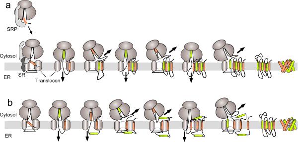
The membrane protein-folding problem can be articulated by two central questions. How is protein topology established by selective peptide transport to opposite sides of the cellular membrane? And how are transmembrane segments inserted, integrated and folded within the lipid bilayer? In eukaryotes, this process usually takes place in the endoplasmic reticulum, coincident with protein synthesis, and is facilitated by the translating ribosome and the Sec61 translocon complex (RTC). At its core, the RTC forms a dynamic pathway through which the elongating nascent polypeptide moves as it is delivered into the cytosolic, lumenal and lipid compartments. This Perspective will focus on emerging evidence that the RTC functions as a protein-folding machine that restricts conformational space by establishing transmembrane topology and yet provides a permissive environment that enables nascent transmembrane domains to efficiently progress down their folding energy landscape.


Figures

Figure 1
Figure 1
Models of polytopic protein biogenesis. (a) Cotranslational biogenesis is initiated as signal recognition particle (SRP) interacts with a signal sequence, binds its receptor (SR) at the ER membrane, and transfers the ribosome nascent chain complex to the Sec61 translocon. Signal sequences (tan cylinders) stimulate ribosome binding and open the translocon pore to initiate peptide movement into the ER lumen. Stop transfer sequences (green cylinders) terminate translocation and redirect the elongating nascent chain beneath the ribosome and into the cytosol. Each sequential TM therefore alters the direction of nascent chain movement through the RTC (bold arrow) to establish transmembrane topology from N- to C-terminus one helix at a time. (b) During AQP1 biogenesis, TM2 fails to terminate translocation, TM3 is initially inserted into the translocon in a type I topology, and TM4 transiently resides on the cytosolic face of the membrane. This sequence of events generates a four-spanning intermediate that is converted to a six spanning topology during or after synthesis of TMs 5–6.
Figure 2
Figure 2
Mechanism of TM integration. (a) During membrane protein biogenesis, TMs are laterally transferred from the proteinaceous environment of the translocon into the lipid bilayer. This may occur in a sequential fashion as each TM is synthesized, in pairs, or in groups. (b)The timing of TM integration will determine, in part, whether helical packing takes place primarily in a lipid or proteinaceous environment. Two potential folding models are illustrated for AQP1. If TMs are sequentially released from the translocon into bulk lipid, then topological maturation of TMs 2–4 will involve spontaneous TM insertion, rotation, and movement of two hydrophilic loops across the lipid bilayer. Alternatively, retention of TMs within or in close proximity to translocon proteins (bottom panel) could potentially reduce energy barriers for peptide transfer and topological maturation.
Figure 3
Figure 3
Potential arrangement of Sec61αβγ heterotrimers (gray cylinders) in the assembled translocon (purple disc) and implications for cotranslational folding. Cryo-EM of empty, solubilized mammalian ER RTCs suggest Sec61 may be present beneath the ribosome in a single copy (a) or a back-to-back tetramer configuration, (b). Both models propose that only one Sec61 protein is used for translocation which provides TM helices (orange and green cylinders) with a single lateral exit site to the translocon periphery. Alternative orientations include a front-to-front Sec61 dimer configuration observed in CryoEM structures of the E. coli SecYEG complex (c), a large central pore derived from fluorescence quenching experiments of functionally intact ER translocons and supported by early low resolution EM studies,, (d), and a related but hypothetical oligomeric front-to-front configuration in which TMs could initially exit Sec61 into the translocon interior (e). These latter models provide a potential means to accommodate multiple helices during translocation and prior to nascent chain movement between subunits into the bilayer.

References

    1. Popot J, Engelman D. Helical membrane protein folding, stability and evolution. Ann. Rev. Biochem. 2000;69:881–922. - PubMed
    1. Bowie J. Solving the membrane protein folding problem. Nature. 2005;438:581–589. - PubMed
    1. Johnson A, van Waes M. The Translocon: A dynamic gateway at the ER membrane. Ann. Rev. Cell Dev. Biol. 1999;15:799–842. - PubMed
    1. Rapoport T, Goder V, Heinrich S, Matlack K. Membrane-protein integration and the role of the translocation channel. Trends Cell Biol. 2004;14:568–575. - PubMed
    1. Blobel G. Intracellular protein topogenesis. Proc. Natl. Acad. Sci. USA. 1980;77:1496–1500. - PMC - PubMed

Publication types

MeSH terms