. 2009 Jun 3;4(6):e5635.
doi: 10.1371/journal.pone.0005635.

Evidence of molecular evolution driven by recombination events influencing tropism in a novel human adenovirus that causes epidemic keratoconjunctivitis

Affiliations

Evidence of molecular evolution driven by recombination events influencing tropism in a novel human adenovirus that causes epidemic keratoconjunctivitis

Michael P Walsh et al. PLoS One. .

Abstract

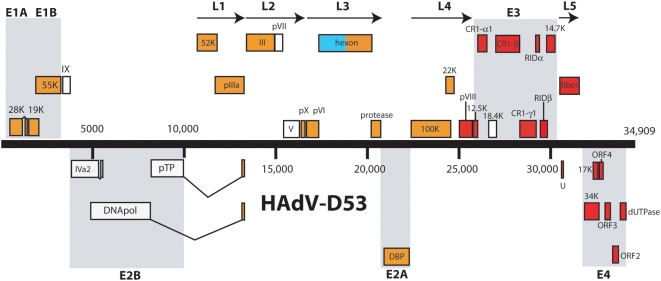
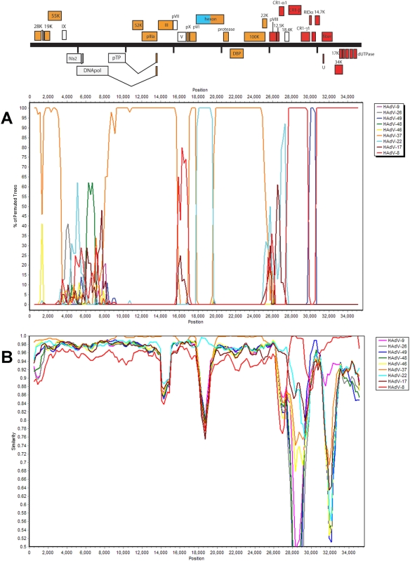
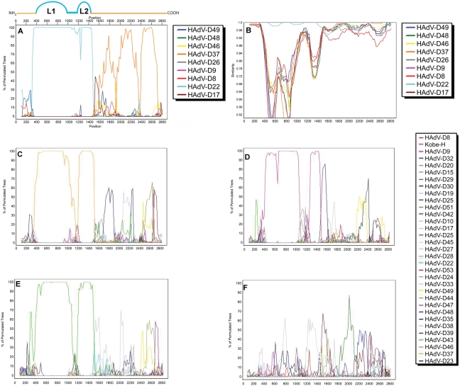
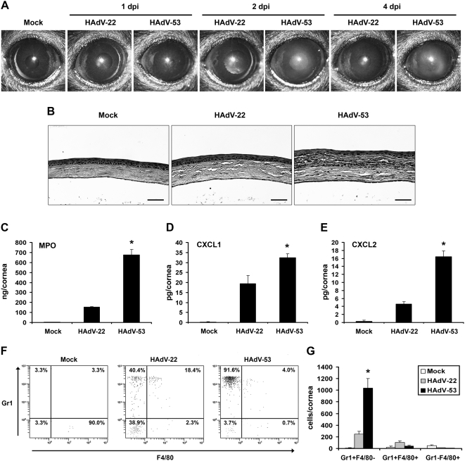
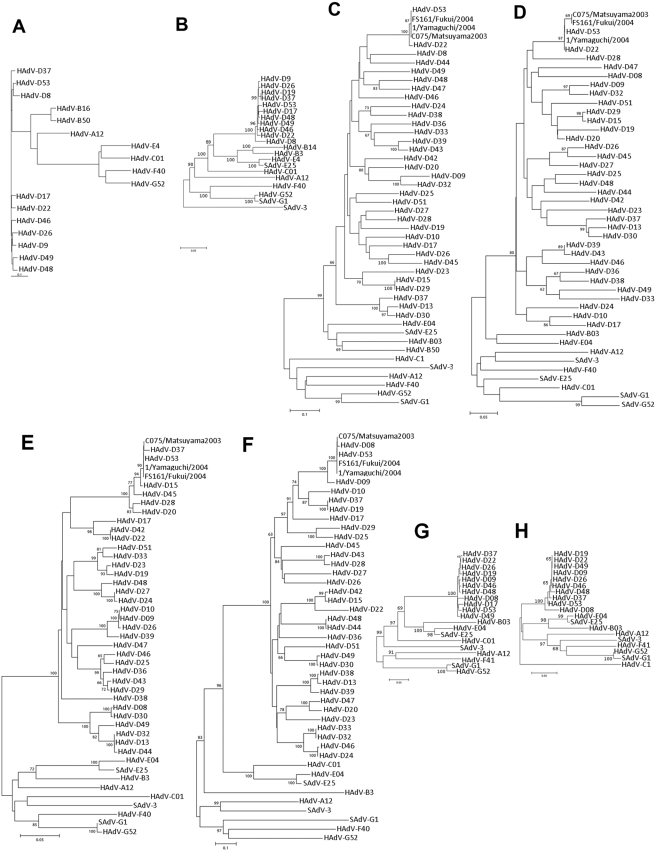
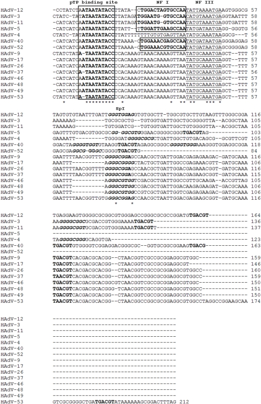
In 2005, a human adenovirus strain (formerly known as HAdV-D22/H8 but renamed here HAdV-D53) was isolated from an outbreak of epidemic keratoconjunctititis (EKC), a disease that is usually caused by HAdV-D8, -D19, or -D37, not HAdV-D22. To date, a complete change of tropism compared to the prototype has never been observed, although apparent recombinant strains of other viruses from species Human adenovirus D (HAdV-D) have been described. The complete genome of HAdV-D53 was sequenced to elucidate recombination events that lead to the emergence of a viable and highly virulent virus with a modified tropism. Bioinformatic and phylogenetic analyses of this genome demonstrate that this adenovirus is a recombinant of HAdV-D8 (including the fiber gene encoding the primary cellular receptor binding site), HAdV-D22, (the epsilon determinant of the hexon gene), HAdV-D37 (including the penton base gene encoding the secondary cellular receptor binding site), and at least one unknown or unsequenced HAdV-D strain. Bootscanning analysis of the complete genomic sequence of this novel adenovirus, which we have re-named HAdV-D53, indicated at least five recombination events between the aforementioned adenoviruses. Intrahexon recombination sites perfectly framed the epsilon neutralization determinant that was almost identical to the HAdV-D22 prototype. Additional bootscan analysis of all HAdV-D hexon genes revealed recombinations in identical locations in several other adenoviruses. In addition, HAdV-D53 but not HAdV-D22 induced corneal inflammation in a mouse model. Serological analysis confirmed previous results and demonstrated that HAdV-D53 has a neutralization profile representative of the epsilon determinant of its hexon (HAdV-D22) and the fiber (HAdV-D8) proteins. Our recombinant hexon sequence is almost identical to the hexon sequences of the HAdV-D strain causing EKC outbreaks in Japan, suggesting that HAdV-D53 is pandemic as an emerging EKC agent. This documents the first genomic, bioinformatic, and biological descriptions of the molecular evolution events engendering an emerging pathogenic adenovirus.


Conflict of interest statement

Competing Interests: The authors have declared that no competing interests exist.

Figures

Figure 1
Figure 1. Genome organization of HAdV-D53.
Genome is represented by a central black horizontal line marked at 5-kbp intervals. Protein-encoding regions are shown as boxes. Boxes above the black line represent open reading frames (ORFs) that are encoded on the forward (or upper) strand. Boxes underneath the black line represent ORFs that are encoded on the reverse (or lower) strand. The colors of the boxes correspond to which adenovirus the protein is most likely descended from: red – HAdV-D8, aqua – HAdV-22, orange – HAdV-D37, white – dissimilar to all known adenoviruses.
Figure 2
Figure 2. Whole genome (A) bootscan and (B) simplot of HAdV-D53 compared to fully sequenced HAdV-D genomes.
Figure 3
Figure 3. Bootscan of HAdV-D species hexon genes demonstrating recombination events.
Comparison of HAdV-D53 by (A) bootscan and (B) simplot with the HAdV-D types which have a fully sequenced genome. (C) HAdV-D13, (D) HAdV-D32, (E) HAdV-D39, and (F) HAdV-D49 were compared to all hexon genes in species HAdV-D by bootscan analysis.
Figure 4
Figure 4. HAdV-D53 induces keratitis.
(A) Clinical appearance of HAdV-D53 keratitis. Virus-free buffer (mock), 104 TCID of HAdV-D22, or HAdV-D53 was injected in the corneal stroma of C57BL/6 mice (n = 8 corneas/group). Corneas were examined under a surgical microscope up to 4 days post-infection. One representative picture from each group is shown at the indicated time points. (B) Histopathology of HAdV-D53 keratitis. Representative histopathological sections at 4 days post-infection of mouse corneas injected with buffer, HAdV-D22, or HAdV-D53 are shown (hematoxylin and eosin stain; scale bar = 50 µ). (C) Myeloperoxidase (MPO) expression in HAdV-D53 keratitis. Mock, HAdV-D22, and HAdV-D53 infected corneas were analyzed by ELISA at 24 hours post-infection for the expression of myeloperoxidase enzyme. (D, E) Chemokine expression in HAdV-D53 keratitis. Expression of CXCL1 (D) and CXCL2 (E) in mock, HAdV-D22, and HAdV-D53 infected corneas were analyzed by ELISA at 16 hpi. Data is mean±SEM from three individual experiments (n = 9 corneas/group). (F, G) Phenotypic analysis of inflammatory cells in HAdV-D53 keratitis. Mock, HAdV-D22, and HAdV-D53 infected corneas at 24 hours post-infection were homogenized and single cell preparations were stained with anti-CD45, anti-Gr1, and anti-F4/80 antibodies. Cells were gated on CD45-positive staining. (F) Representative dot plots or (G) quantification of three separate experiments is shown for each group (mean cells/cornea±SEM, n = 9 corneas/group). In all experiments statistical significance is denoted by *, P<.05 as determined by ANOVA with Scheffe's multiple comparison test.
Figure 5
Figure 5. Phylogenetic analysis of HAdV-D53.
Analysis of HAdV-D53 is based on the nucleic acid sequence of (A) complete genomes, as well as the predicted amino acid sequences of (B) polymerase, (C) L1 and (D) L2 of the hexon protein penton, (E) β-determinant, (F) γ-determinant, (G) pV and (H) pVII. Numbers denote human adenovirus serotypes. HAdV-D53 (in bold) shows the new isolate. The numbers close to the nodes represents bootstrap pseudoreplicates.
Figure 6
Figure 6. Analysis of the HAdV-D53 inverted terminal repeat (ITR).
NF I, NF III, SpI, and pTP binding motifs are marked. The ATF binding site is TGACGT.

References

    1. Gordon YJ, Aoki K, Kinchington PR. Adenovirus keratoconjunctivitis. In: Pepose JS, Holland GN, Wilhelmus KR, editors. Ocular Infection and Immunity. St. Louis: Mosby; 1996. pp. 877–94.
    1. Rowe WP, Huebner RJ, Gilmore LK, Parrott RH, Ward TG. Isolation of a cytopathogenic agent from human adenoids undergoing spontaneous degeneration in tissue culture. Proc Soc Exp Biol Med. 1953;84:570–573. - PubMed
    1. Hilleman MR, Werner JH. Recovery of new agent from patients with acute respiratory illness. Proc Soc Exp Biol Med. 1954;851:183–188. - PubMed
    1. Ginsberg HS, Gold E, Jordan WS, Jr, Katz S, Badger GF, Dingle JH. Relation of the new respiratory agents to acute respiratory diseases. Am J Pub Health. 1955;45:915–922. - PMC - PubMed
    1. Dingle JH, Langmuir AD. Epidemiology of acute, respiratory disease in military recruits. Am Rev Respir Dis. 1968;976(Suppl):1–65. - PubMed

Publication types