. 2009 May 28;4(5):e5727.
doi: 10.1371/journal.pone.0005727.

Conformational targeting of fibrillar polyglutamine proteins in live cells escalates aggregation and cytotoxicity

Affiliations

Conformational targeting of fibrillar polyglutamine proteins in live cells escalates aggregation and cytotoxicity

Erik Kvam et al. PLoS One. .

Abstract

Background: Misfolding- and aggregation-prone proteins underlying Parkinson's, Huntington's and Machado-Joseph diseases, namely alpha-synuclein, huntingtin, and ataxin-3 respectively, adopt numerous intracellular conformations during pathogenesis, including globular intermediates and insoluble amyloid-like fibrils. Such conformational diversity has complicated research into amyloid-associated intracellular dysfunction and neurodegeneration. To this end, recombinant single-chain Fv antibodies (scFvs) are compelling molecular tools that can be selected against specific protein conformations, and expressed inside cells as intrabodies, for investigative and therapeutic purposes.

Methodology/principal findings: Using atomic force microscopy (AFM) and live-cell fluorescence microscopy, we report that a human scFv selected against the fibrillar form of alpha-synuclein targets isomorphic conformations of misfolded polyglutamine proteins. When expressed in the cytoplasm of striatal cells, this conformation-specific intrabody co-localizes with intracellular aggregates of misfolded ataxin-3 and a pathological fragment of huntingtin, and enhances the aggregation propensity of both disease-linked polyglutamine proteins. Using this intrabody as a tool for modulating the kinetics of amyloidogenesis, we show that escalating aggregate formation of a pathologic huntingtin fragment is not cytoprotective in striatal cells, but rather heightens oxidative stress and cell death as detected by flow cytometry. Instead, cellular protection is achieved by suppressing aggregation using a previously described intrabody that binds to the amyloidogenic N-terminus of huntingtin. Analogous cytotoxic results are observed following conformational targeting of normal or polyglutamine-expanded human ataxin-3, which partially aggregate through non-polyglutamine domains.

Conclusions/significance: These findings validate that the rate of aggregation modulates polyglutamine-mediated intracellular dysfunction, and caution that molecules designed to specifically hasten aggregation may be detrimental as therapies for polyglutamine disorders. Moreover, our findings introduce a novel antibody-based tool that, as a consequence of its general specificity for fibrillar conformations and its ability to function intracellularly, offers broad research potential for a variety of human amyloid diseases.


Conflict of interest statement

Competing Interests: The authors have declared that no competing interests exist.

Figures

Figure 1
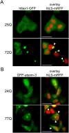
Figure 1. Localization of GFP-tagged httex1 and full-length ataxin-3 in live ST14A striatal progenitor cells.
ST14A cells were transiently transfected with httex1-GFP or GFP-ataxin-3 reporters containing normal or disease-associated polyglutamine tract lengths, and NLS-mRFP to label nuclei. Representative live-cell images were captured 48 hours post-transfection as described in Materials and Methods. (A) GFP-tagged httex1-25Q localizes diffusely in the cytoplasm while mutant httex1-72Q precipitously forms large aggregates (arrows) in the cytoplasm, which often appear juxtaposed to nuclei. (B) GFP-tagged ataxin-3-24Q localizes diffusely within ST14A cells while mutant ataxin-3-77Q forms small aggregates (arrows) predominately in the cytoplasm. Scale bars, 25 µm.
Figure 2
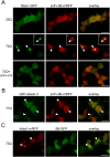
Figure 2. scFv-6E fluorobodies co-localize with intracellular aggregates of mutant httex1 and ataxin-3 in live cells.
ST14A cells were transiently transfected with the indicated GFP- and mRFP-tagged reporters, and representative live-cell images were captured 48 hours post-transfection as described in Materials and Methods. (A) mRFP-tagged scFv-6E fluorobody co-localizes as intracellular puncta with cytoplasmic aggregates of GFP-tagged httex1-72Q (arrows), occasionally in a ring-like pattern (inset i). Transfection with unlabeled scFv-C4 efficiently suppresses co-localization of scFv-6E fluorobody with inclusions by inhibiting the aggregation of httex1-72Q-GFP. In the absence of httex1 inclusions, scFv-6E fluorobody exhibits a diffuse localization pattern. (B) mRFP-tagged scFv-6E fluorobody co-localizes with cytoplasmic and nuclear aggregates of GFP-tagged ataxin-3-77Q (arrows). (C) GFP-labeled control intrabody (B8) selected against botulinum neurotoxin light chain protease fails to co-localize with mutant httex1-72Q-mRFP inclusions. Scale bars, 25 µm.
Figure 3
Figure 3. NLS-fused scFv-6E intrabody retargets polyglutamine aggregates but not soluble polyglutamine species in situ.
Antibody-antigen interactions were probed in ST14A cells using NLS-fused scFv-6E to sequester both intrabody and bound antigen into nuclei. NLS-mRFP fluorescence marks nuclei. Representative live-cell images were captured as described in Materials and Methods. (A) NLS-fused scFv-6E sequesters small cytoplasmic aggregates of GFP-labeled httex1-72Q (inset, arrow) but not soluble species into nuclei. In contrast, a control NLS-fused intrabody (scFv-C4-NLS) completely recruits soluble httex1-Q72-GFP species into nuclei from the cytoplasm. (B) NLS-fused scFv-6E completely sequesters aggregates of GFP-labeled ataxin-3-77Q (inset, arrow) but not soluble species into nuclei. Scale bars, 25 µm.
Figure 4
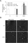
Figure 4. scFv-6E enhances the fibrillar aggregation of mutant httex1 in solution.
(A) Fluorescence intensity of Thioflavine S-stained httex1-51Q fibrils formed in the presence or absence of scFv antibodies. Purified httex1-51Q, scFv-6E, and scFv-αPLB proteins were prepared as described in Materials and Methods. Samples were analyzed 0, 3, and 24 hours after initiating aggregation. Each bar represents mean Thioflavine S fluorescence (arbitrary fluorescence units, AFU) from reactions performed in triplicate. Statistical significance (** p<0.01) was determined by ANOVA. (B) Representative AFM images of httex1-51Q aggregation reactions conducted in the absence or presence of scFv-6E. AFM scan size, 5 µm×5 µm.
Figure 5
Figure 5. scFv-6E intrabody enhances mutant httex1 aggregation in ST14A cells.
(A) Representative live-cell images depicting the localization patterns of httex1-72Q-GFP and NLS-mRFP in the absence or presence of intrabodies. scFv-6E intrabody increases the frequency of cytoplasmic httex1-72Q aggregates while scFv-C4 intrabody blocks aggregate formation in ST14A cells. Asterisks denote cells where NLS-mRFP nuclear import is impaired, indicating cell stress. Scale bars, 25 µm. (B) Relative aggregation propensity of httex1-72Q in the absence or presence of intrabodies. Aggregates of httex1-72Q-GFP were scored as described in Materials and Methods. Statistical significance (*** p<0.0001) was determined by ANOVA (n = 4). (C) FACS histogram of httex1-72Q-GFP fluorescence intensity (arbitrary fluorescence units, AFU) plotted as a function of cell number. ST14A cells co-expressing httex1-72Q-GFP and either scFv-6E (black) or empty vector (gray) were sorted by fluorescence intensity as described in Materials and Methods. Representative data from triplicate experiments were superimposed.
Figure 6
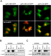
Figure 6. Relative intrabody solubility.
(A) Live images of ST14A cells co-transfected with GFP-tagged fluorobodies and NLS-mRFP, a nuclear marker. Cells were imaged 48 hours post-transfection as described in Materials and Methods. scFv-6E fluorobody predominately localizes as a diffuse soluble protein similar to anti-huntingtin scFv-C4 fluorobody. scFv-ML3.9 fluorobody localizes almost entirely as inclusions. Note NLS-mRFP nuclear import is perturbed in the presence of scFv-ML3.9 inclusions (asterisks), indicating cell stress. (B) Biochemical fractionation of HA-tagged fluorobodies from ST14A cells. Detergent-soluble and -insoluble cell lysates were prepared as described in Materials and Methods. (C) Biochemical fraction of HA-tagged intrabodies following elevated overexpression. Detergent-soluble and -insoluble cell lysates were prepared as described in Materials and Methods.
Figure 7
Figure 7. scFv-6E and -C4 intrabodies differentially affect mutant httex1-mediated toxicity and oxidative stress in situ.
(A) Two-color FACS dot-plots from representative experiments in which ST14A cells were transiently transfected with the indicated reporters, stained with propidium iodide (PI) to label dead cells, and co-sorted for GFP and PI fluorescence intensity. A total of 30,000 cells were plotted on a logarithmic scale of arbitrary fluorescence intensity. Grid lines were positioned after calibrating the flow cytometer as described in Materials and Methods. Lower right quadrants demarcate viable transfected cells (GFP+ PI). Upper right quadrants signify PI-labeled dead or dying transfected cells (GFP+ PI+). Note that scFv-6E intrabody shifts httex1-72Q-GFP fluorescence to a higher intensity among PI-labeled cells (arrows), indicating that these cells died with intensely fluorescent aggregates. (B) Percentage of cell death observed by two-color FACS analysis with PI. Cell death was calculated by dividing the number of PI-stained transfected cells (GFP+ PI+, see upper right quadrants in A) over the total number of transfected cells (GFP+ PI+ and GFP+ PI, see upper and lower right quadrants in A). Statistical significance from httex1-72Q-GFP alone (** p<0.001, *** p<0.0001) was determined by ANOVA (n = 3). (C) Relative cell staining of PI, YOPRO-1, and dihydroethidium (DHE) dyes as detected by two-color FACS. ST14A cells were transiently transfected with the indicated GFP- or mRFP-labeled httex1 reporters (with or without scFv-6E and scFv-C4 intrabodies) and incubated with PI, YOPRO-1, or DHE as described in Materials and Methods. Cell staining percentages were calculated by dividing the number of dye-stained transfected cells by the total number of transfected cells, and data were normalized to non-expanded httex1-25Q alone. Statistical significance from expanded httex1-72Q alone (* p<0.01, ** p<0.001, *** p<0.0001) was determined by ANOVA (n = 3).
Figure 8
Figure 8. scFv-6E intrabody enhances aggregation and toxicity of both normal and disease-associated ataxin-3.
(A) Live-cell images depicting GFP localization patterns of non-disease-associated ataxin3-24Q, ataxin3-ΔQ (lacking a polyglutamine tract), or httex1-25Q in the absence or presence of scFv-6E intrabody. scFv-6E intrabody stimulates the intracellular aggregation of ataxin-3 (arrows) but not httex1-25Q. Scale bars, 25 µm. (B) Relative aggregation propensity of normal (24Q) or disease-linked (77Q) ataxin-3 in the absence or presence of scFv-6E intrabody. GFP-tagged ataxin-3 aggregates were scored in ST14A as described in Materials and Methods. Statistical significance (** p<0.001, *** p<0.0001) was determined by ANOVA (n = 3). (C) Relative cell staining of propidium iodide (PI) and dihydroethidium (DHE) vital dyes as detected by two-color FACS. ST14A cells were transiently transfected with normal (24Q) or disease-linked (77Q) GFP-ataxin-3 and either scFv-6E intrabody or empty vector. Cell staining percentages were calculated as described in Materials and Methods, and data were normalized to GFP-ataxin-3-24Q alone. Statistical significance (* p<0.01, *** p<0.0001) from GFP-ataxin-3-24Q was determined by ANOVA (n = 3).

Similar articles

Cited by

References

    1. Uversky VN. Nanoimaging in protein-misfolding and -conformational diseases. Nanomed. 2007;2:615–643. - PubMed
    1. Bates GP. Huntingtin aggregation and toxicity in Huntington's disease. Lancet. 2003;361:1642–1644. - PubMed
    1. Gutekunst CA, Li SH, Yi H, Mulroy JS, Kuemmerle S, et al. Nuclear and neuropil aggregates in Huntington's disease: relationship to neuropathology. J Neurosci. 1999;19:2522–2534. - PMC - PubMed
    1. Weiss A, Klein C, Woodman B, Sathasivam K, Bibel M, et al. Sensitive biochemical aggregate detection reveals aggregation onset before symptom development in cellular and murine models of Huntington's disease. J Neurochem. 2008;104:846–858. - PubMed
    1. Mangiarini L, Sathasivam K, Seller M, Cozens B, Harper A, et al. Exon 1 of the HD gene with an expanded CAG repeat is sufficient to cause a progressive neurological phenotype in transgenic mice. Cell. 1996;87:493–506. - PubMed

Publication types

MeSH terms