

Common features of megakaryocytes and hematopoietic stem cells: what's the connection?
- PMID: 19492306
- PMCID: PMC2741141
- DOI: 10.1002/jcb.22184
Common features of megakaryocytes and hematopoietic stem cells: what's the connection?
Abstract
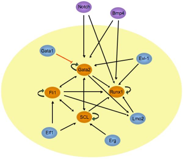
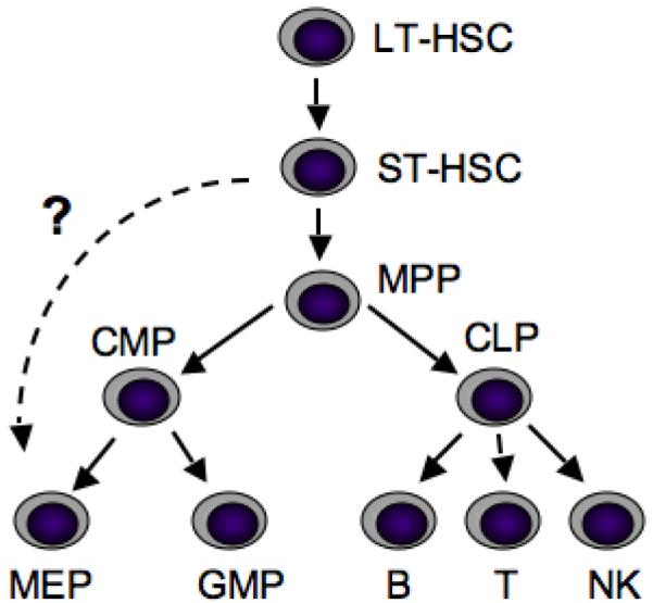
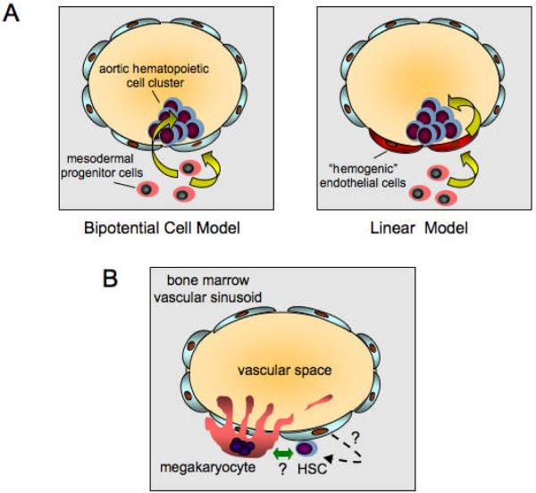
Megakaryocytes (Mks) are rare polyploid bone marrow cells whose function is to produce blood platelets. Since the purification and cloning of the major Mk cytokine, thrombopoietin, in 1994, considerable progress has been made in understanding the biology of Mk development. Remarkably, these advances have revealed a number of key features of Mks that are shared with hematopoietic stem cells (HSCs), such as common surface receptors, lineage-specific transcription factors, and specialized signaling pathways. Why there should be such a close connection between these two cell types remains unclear. In this Prospect article, we summarize the data supporting these shared features and speculate on possible teleological bases. In particular, we focus on common links involving developmental hierarchy, endothelial cells, and bone marrow niche interactions. This discussion highlights new data showing close ontologic relationship between HSCs and specialized "hemogenic" endothelial cells during development, and functional overlap between Mks/platelets and endothelial cells. Overall, these findings may be of relevance in the development of techniques for HSC ex vivo culture and/or possible generation of HSCs via somatic cell reprogramming.
(c) 2009 Wiley-Liss, Inc.
Figures
References
-
- Adolfsson J, Mansson R, Buza-Vidas N, Hultquist A, Liuba K, Jensen CT, Bryder D, Yang L, Borge OJ, Thoren LA, Anderson K, Sitnicka E, Sasaki Y, Sigvardsson M, Jacobsen SE. Identification of Flt3+ lympho-myeloid stem cells lacking erythro-megakaryocytic potential a revised road map for adult blood lineage commitment. Cell. 2005;121:295–306. - PubMed
-
- Argiropoulos B, Humphries RK. Hox genes in hematopoiesis and leukemogenesis. Oncogene. 2007;26:6766–76. - PubMed
-
- Avecilla ST, Hattori K, Heissig B, Tejada R, Liao F, Shido K, Jin DK, Dias S, Zhang F, Hartman TE, Hackett NR, Crystal RG, Witte L, Hicklin DJ, Bohlen P, Eaton D, Lyden D, de Sauvage F, Rafii S. Chemokine-mediated interaction of hematopoietic progenitors with the bone marrow vascular niche is required for thrombopoiesis. Nat Med. 2004;10:64–71. - PubMed
-
- Azcoitia V, Aracil M, Martinez AC, Torres M. The homeodomain protein Meis1 is essential for definitive hematopoiesis and vascular patterning in the mouse embryo. Dev Biol. 2005;280:307–20. - PubMed
-
- Ben-David Y, Giddens EB, Letwin K, Bernstein A. Erythroleukemia induction by Friend murine leukemia virus: insertional activation of a new member of the ets gene family, Fli-1, closely linked to c-ets-1. Genes Dev. 1991;5:908–18. - PubMed
Publication types
MeSH terms
Grants and funding
LinkOut - more resources
Full Text Sources
Other Literature Sources
Medical
