Review
. 2009;48(30):5406-15.
doi: 10.1002/anie.200805179.

Engineering substrate topography at the micro- and nanoscale to control cell function

Affiliations
Review

Engineering substrate topography at the micro- and nanoscale to control cell function

Christopher J Bettinger et al. Angew Chem Int Ed Engl. 2009.

Abstract

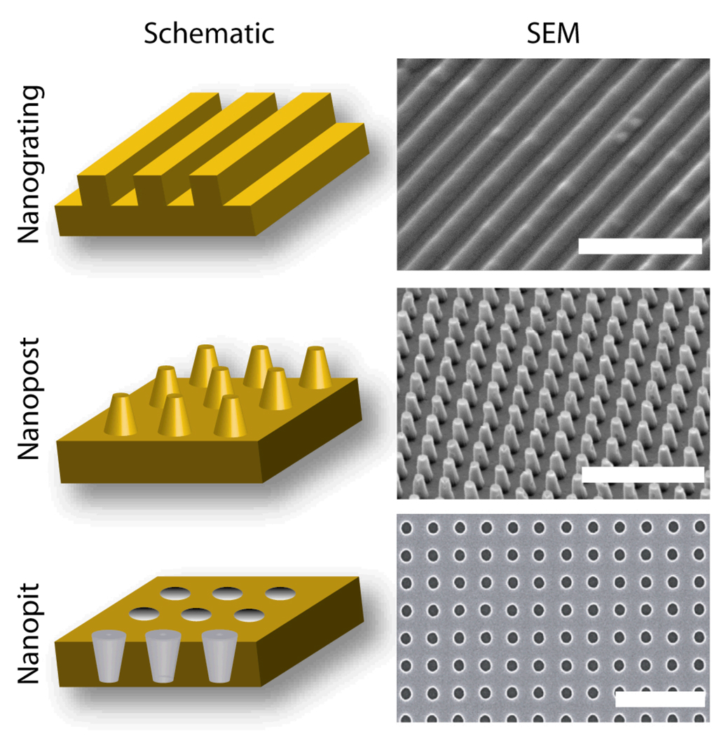
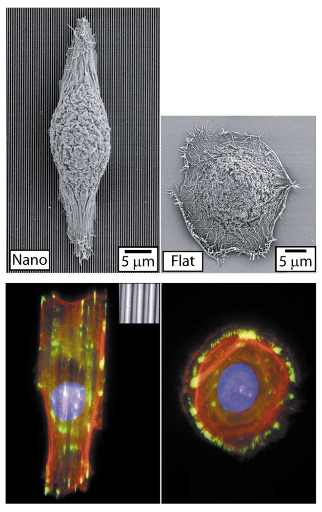
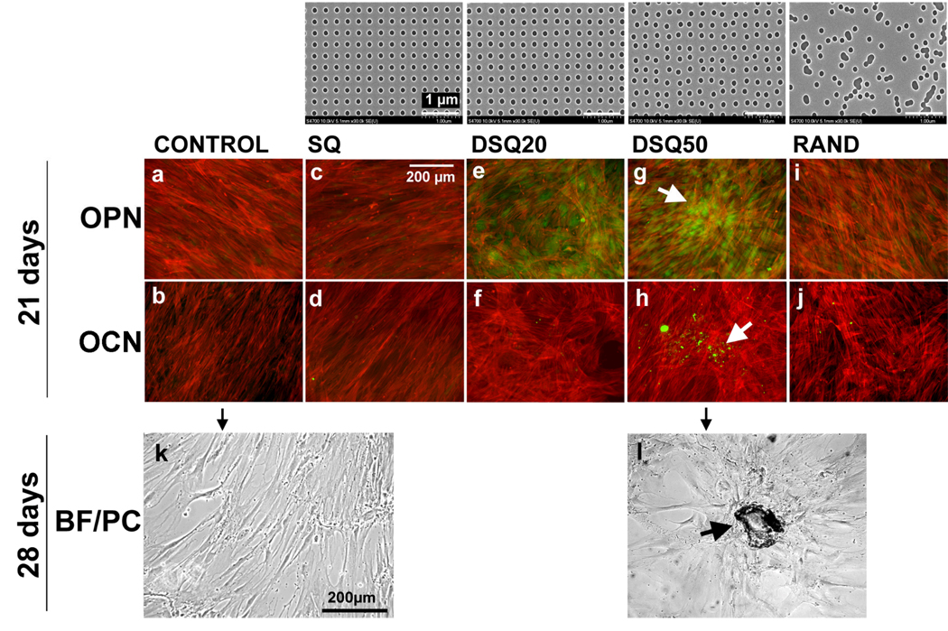
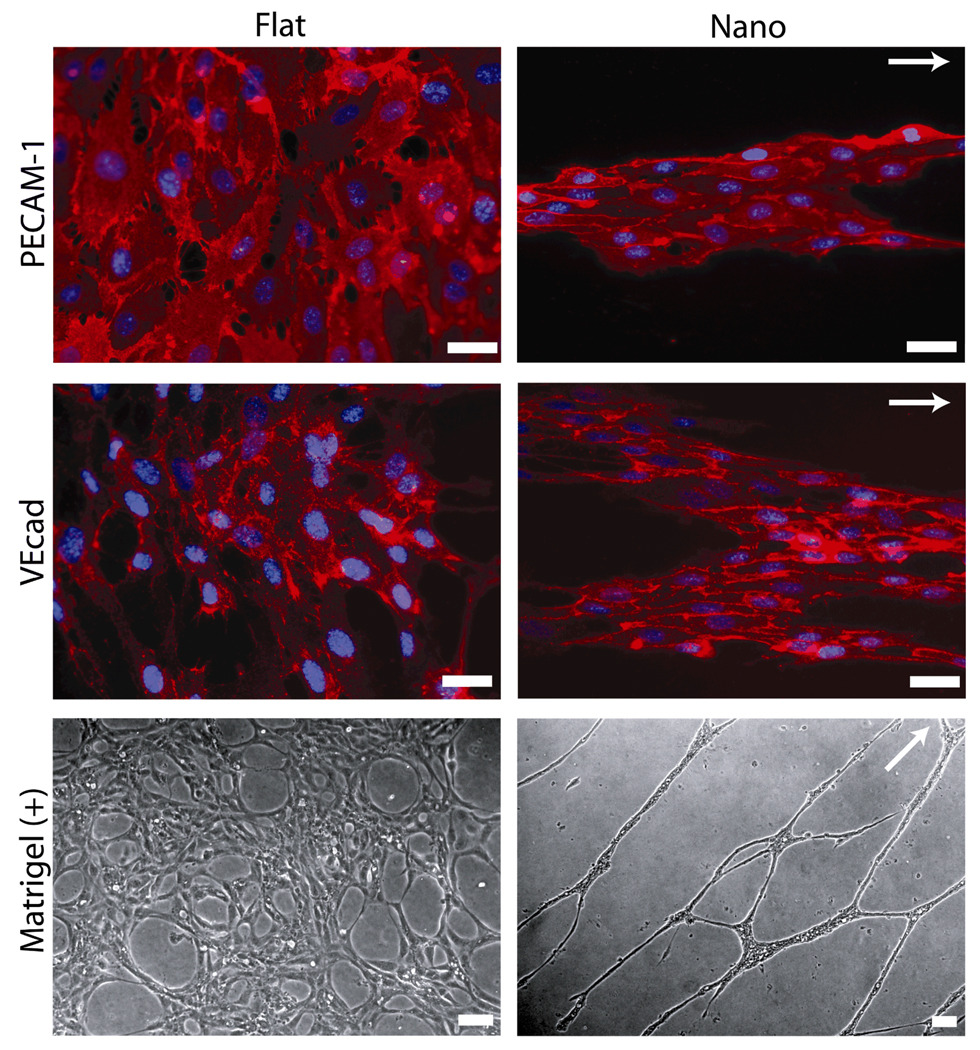
The interaction of mammalian cells with nanoscale topography has proven to be an important signaling modality in controlling cell function. Naturally occurring nanotopographic structures within the extracellular matrix present surrounding cells with mechanotransductive cues that influence local migration, cell polarization, and other functions. Synthetically nanofabricated topography can also influence cell morphology, alignment, adhesion, migration, proliferation, and cytoskeleton organization. We review the use of in vitro synthetic cell-nanotopography interactions to control cell behavior and influence complex cellular processes, including stem-cell differentiation and tissue organization. Future challenges and opportunities in cell-nanotopography engineering are also discussed, including the elucidation of mechanisms and applications in tissue engineering.


Figures

Figure 1
Figure 1
Schematics and SEM Images of Representative Nanotopography Geometries. Three basic nanotopography geometries include nanograting (45° tilt, scale bar represents 5 µm), nanopost array (15° tilt, scale bar represents 5 µm) and nanopit array (0° tilt, scale bar represents 1 µm). Schematics not drawn to scale.
Figure 2
Figure 2
Cell-Nanograting Response in Epithelial Cells. Epithelial cells respond to nanograting through alignment and elongation to the grating axis as evident through fluorescent and SEM micrographs. Other cell types exhibit similar morphological responses when cultured on nanograting substrates (Table 1). Reproduced with permission of the Company of Biologists.
Figure 3
Figure 3
Directed Differentiation of Human Mesenchymal Stem Cells to Osteoblast Lineage Using Nanopit Substrates. The top row shows images of nanopit arrays fabricated by electron beam lithography. All have 120-nm-diameter pits (100 nm deep, absolute or average 300 nm centre–centre spacing) with square (SQ), displaced square 20 (±20 nm from true centre, DSQ20), displaced square 50 (±50 nm from true centre, DSQ50) and random placements (RAND). Human MSCs cultured on a planar control substrate (a,f), note the fibroblastic appearance and no osteopontin (OPN) or osteocalcein (OCN) positive cells; on the SQ array (b,g), note the fibroblastic appearance and no OPN or OCN positive cells; on the DSQ20 array (c,h), note OPN positive cells; on the DSQ50 array (d,i), note OPN and OCN positive cells and nodule formation (arrows); on the RAND array (e,j), note the osteoblast morphology, but no OPN or OCN positive cells. (k,l) Phase-contrast/bright-field images showing that hMSCs cultured on the control (k) had a fibroblastic morphology after 28 d, whereas hMSCs cultured on DSQ50 arrays (l) exhibited mature bone nodules containing mineral. Reprinted by permission from Macmillan Publishers Ltd: Nature Materials (M Dalby, N Gadegaard, R Tare, A Andar, M O Riehle, P Herzyk, C D W Wilkinson, R O C Oreffo. Nat Mater 6. 997), copyright (2007).
Figure 4
Figure 4
Nanograting Substrates Promote Organized Multicellular Structures and Enhance In Vitro Capillary Tube Formation. Protein-level expression of platelet/endothelial cell adhesion molecule-1 (PECAM-1) and vascular endothelial cadherin (VEcad), two endothelial cell markers, was constant in endothelial cells cultured on both planar and nanograting substrates. However, endothelial cells cultured on planar substrates formed confluent monolayers while endothelial cells cultured on nanogratings were organized into multicellular band structures. These aligned band structures formed aligned capillary tubes in an in vitro matrigel assay (Matrigel (+)). The grating axis is indicated by the white arrows. Scale bars represent 50 µm. Adapted from C J Bettinger, Z Zhang, S Gerecht, J T Borenstein, R Langer: Enhancement of In Vitro Capillary Tube Formation by Substrate Nanotopography. Adv Mater. 2008. 20. 99–103. Copyright Wiley-VCH Verlag GmbH & Co. KGaA. Reproduced with permission.

Similar articles

Cited by

References

    1. Goodman SL, Sims PA, Albrecht RM. Biomaterials. 1996;17:2087. - PubMed
    1. Abrams GA, Goodman SL, Nealey PF, Franco M, Murphy CJ. Cell Tissue Res. 2000;299:39. - PubMed
    1. Pamula E, De Cupere V, Dufrene YF, Rouxhet PG. J Colloid Interf Sci. 2004;271:80. - PubMed
    1. Bozec L, van der Heijden G, Horton M. Biophys J. 2007;92:70. - PMC - PubMed
    1. Wolf K, Muller R, Borgmann S, Brocker EB, Friedl P. Blood. 2003;102:3262. - PubMed

Publication types

LinkOut - more resources