. 2009 Jul 17;4(7):547-56.
doi: 10.1021/cb900062k.

An in vivo covalent TMP-tag based on proximity-induced reactivity

Affiliations

An in vivo covalent TMP-tag based on proximity-induced reactivity

Sarah S Gallagher et al. ACS Chem Biol. .

Abstract

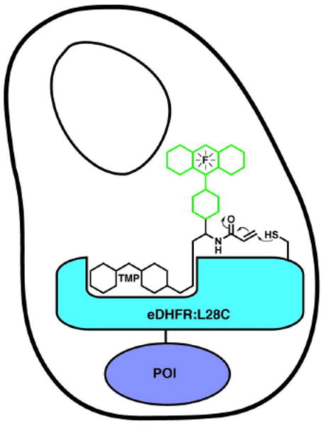
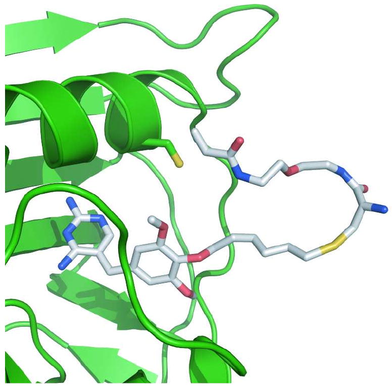
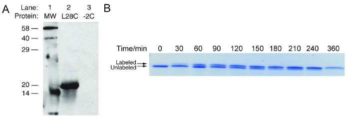
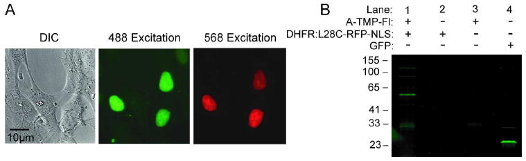
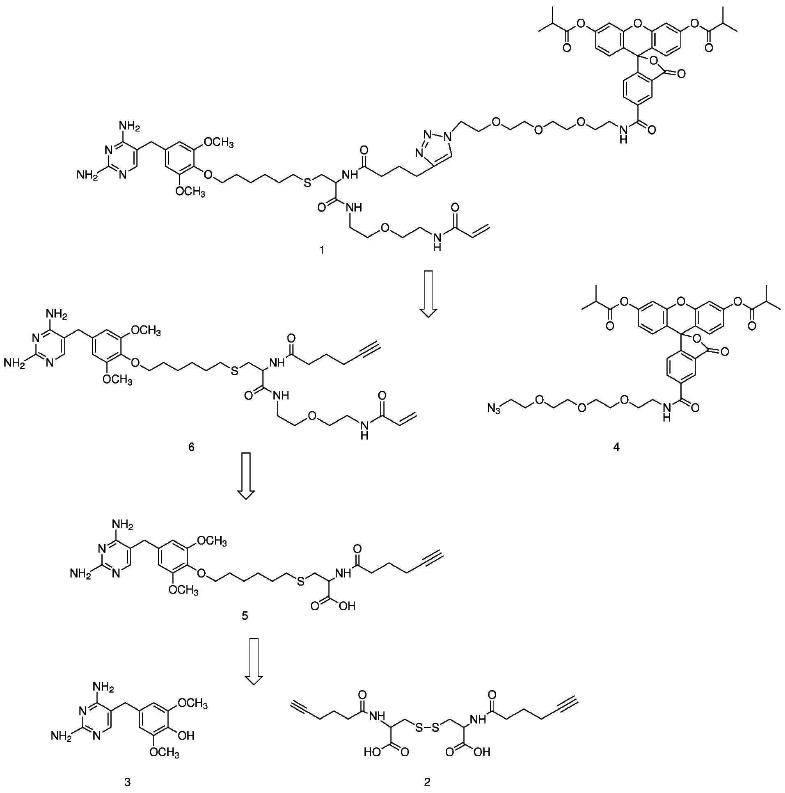
Chemical tags for live cell imaging are emerging as viable alternatives to the fluorescent proteins for labeling proteins with small molecule probes. Among reported chemical tags, trimethoprim (TMP)-tag stands out for having sufficient cell permeability and selectivity to allow imaging of intracellular proteins. TMP-tag provides a noncovalent label in which the protein of interest is fused to E. coli dihydrofolate reductase (DHFR) and then labeled with a cell-permeable TMP-probe heterodimer. To complement the utility of the noncovalent TMP-tag, we sought to render the TMP-tag covalent for applications such as single-molecule tracking and pulse-chase labeling that would benefit from a more permanent modification. On the basis of the long-standing use of proximity-induced reactivity for irreversible inhibitor design and its more recent application to in vitro chemical biology tools, we designed an eDHFR variant with a unique cysteine residue positioned to react with an acrylamide electrophile installed on the TMP-probe label. In vitro experiments show that the eDHFR:L28C nucleophile reacts rapidly and quantitatively with the TMP-acrylamide-probe. Most significantly, the balance in reactivity provided by the acrylamide electrophile allows intracellular proteins tagged with eDHFR:L28C to be labeled with a TMP-acrylamide-fluorescein heterotrimer in live cells with minimal background. Thus, the TMP electrophile described here can be used immediately as a covalent chemical tag in live cells. Moreover, proximity-induced reactivity is shown to be sufficiently selective for use in a living cell, suggesting a general approach for the development of orthogonal covalent chemical tags from existing noncovalent ligand-protein pairs.


Figures

Figure 1
Figure 1
A covalent TMP-tag. We sought to convert our non-covalent trimethoprim (TMP) E. coli dihydrofolate reductase (DHFR) chemical tag into a covalent tag using proximity-induced reactivity. An acrylamide electrophile was installed on the trimethoprim-fluorophore (TMP-F) label. Then molecular modeling was used to design a Cys nucleophile on the surface of eDHFR optimally positioned to react with the acrylamide electrophile when TMP bound to the active-site of eDHFR. A covalent TMP-tag would be advantageous for pulse-chase experiments, single-molecule imaging and other applications that benefit from a more permanent chemical modification.
Figure 2
Figure 2
The design of a covalent TMP-DHFR pair. Cartoon of a TMP-acrylamide heterodimer bound to eDHFR, with an engineered Leu28Cys in position to react with the acrylamide electrophile. The eDHFR protein is represented in green as a ribbon diagram, with the Cys28 residue shown using the stick representation. The TMP-acrylamide heterodimer is also represented as sticks with coloring based on elements. Since no structure has been solved of TMP bound to eDHFR, the model was created by structural alignment of eDHFR (22) with the L. casei DHFR (21). The A-TMP structure was created in MacroModel (23) and then superimposed on TMP in the eDHFR model. The graphic was prepared using PyMOL (24).
Figure 3
Figure 3
Covalent labeling with A-TMP-tag in vitro. To demonstrate the rate and efficiency of covalent labeling, A-TMP-biotin was incubated with purified eDHFR and then analyzed by sodium dodecyl sulfate (SDS)-PAGE. a) Western analysis of in vitro covalent labeling reaction. Purified eDHFR at a concentration of 5 μM was incubated with 10 μM A-TMP-biotin in phosphate buffered saline (PBS) with 1 mM glutathione at 37°C for three hours. A biotinylated protein ladder was run in Lane 1. Lanes 2, 5 μM purified eDHFR:L28C incubated with 10 μM A-TMP-biotin; Lane 3, 5 μM eDHFR:−2C incubated with 10 μM A-TMP-biotin. b) Determination of the rate of covalent labeling. Purified eDHFR:L28C was labeled under the same reaction conditions in a. At various time points, aliquots were quenched with 6× SDS. The reaction was analyzed by SDS-PAGE and Coomassie staining and it was determined that 50% labeling occurs in approximately 50 minutes and that the reaction is near quantitative at the end.
Figure 4
Figure 4
Covalent labeling with A-TMP-tag in vivo. To demonstrate the specificity of covalent labeling in living cells, A-TMP-fluorescein was used to label wild type fibroblasts expressing a nucleus-targeted eDHFR:L28C-RFP fusion protein. a) Live cell imaging of intracellular proteins using the covalent A-TMP-tag. A nucleus-targeted eDHFR:L28C-RFP fusion protein was labeled with A-TMP-fluorescein (A-TMP-Fl) in wild type fibroblasts. Cells transiently transfected with vector encoding the nucleus-targeted eDHFR:L28C-RFP fusion were incubated with 1 μM A-TMP-Fl in phosphate buffered saline (PBS) for ten minutes, washed twice with PBS, incubated for one hour, and then imaged using live cell, confocal microscopy. Confocal micrographs: left column, differential image contrast; middle column, excitation of fluorescein at 488 nm; right column, excitation of RFP at 568 nm. b) In-gel fluorescence scanning analysis of in vivo covalent labeling reaction. Cells were nucleofected 48 hours prior to labeling experiment. The cells were then washed with PBS and incubated for 3 hours in 4 mL of complete DMEM either with or without 1 μM A-TMP-Fl. The cells were washed twice with PBS, trypsinized, and pelleted. The supernatant was removed, and the cells were lysed and the lysate was analyzed by sodium dodecyl sulfate (SDS)-PAGE and in-gel fluorescence scanning, exciting with a 488 nm laser. Lane 1, cells transiently transfected with nucleus-targeted eDHFR:L28C-RFP and incubated with A-TMP-Fl; Lane 2, cells transiently transfected with nucleus-targeted eDHFR:L28C-RFP but not incubated with A-TMP-Fl; Lane 3, untransfected cells incubated with A-TMP-Fl; Lane 4, a positive control of cells transiently transfected with a cytostolic GFP vector. These results show A-TMP-Fl reacts covalently and selectively with eDHFR:L28C in vivo.
Scheme 1
Scheme 1
Retrosynthetic analysis of TMP-fluorophore-acrylamide heterotrimer.

Similar articles

Cited by

References

    1. Giepmans BNG, Adams SR, Ellisman MH, Tsien RY. Review - The fluorescent toolbox for assessing protein location and function. Science. 2006;312:217–224. - PubMed
    1. Tsien RY. The green fluorescent protein. Annual Review of Biochemistry. 1998;67:509–544. - PubMed
    1. Shaner NC, Patterson GH, Davidson MW. Advances in fluorescent protein technology. J Cell Sci. 2007;120:4247–4260. - PubMed
    1. O'Hare HM, Johnsson K, Gautier A. Chemical probes shed light on protein function. Curr Opin Struct Biol. 2007;17:488–494. - PubMed
    1. Miller LW, Cornish VW. Selective chemical labeling of proteins in living cells. Curr Opin Chem Biol. 2005;9:56–61. - PubMed

Publication types

Substances