. 2009 Aug;110(4):1191-202.
doi: 10.1111/j.1471-4159.2009.06202.x. Epub 2009 May 31.

Sphingosine-1-phosphate receptors mediate neuromodulatory functions in the CNS

Affiliations

Sphingosine-1-phosphate receptors mediate neuromodulatory functions in the CNS

Laura J Sim-Selley et al. J Neurochem. 2009 Aug.

Abstract

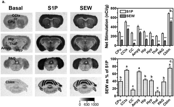
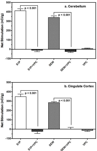
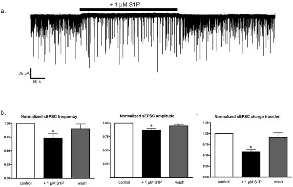
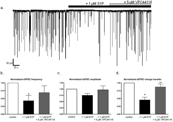
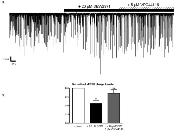
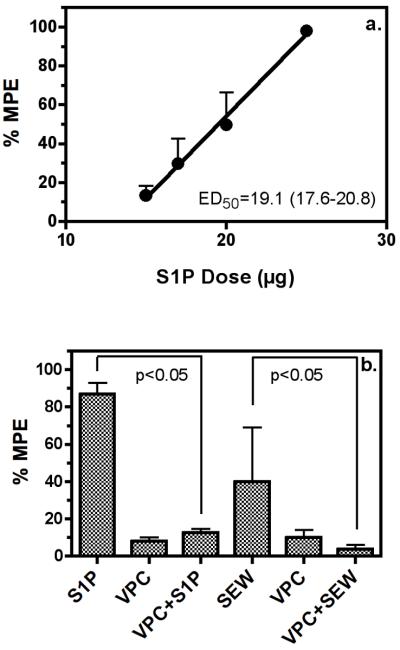
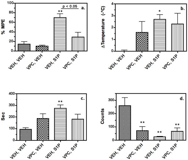
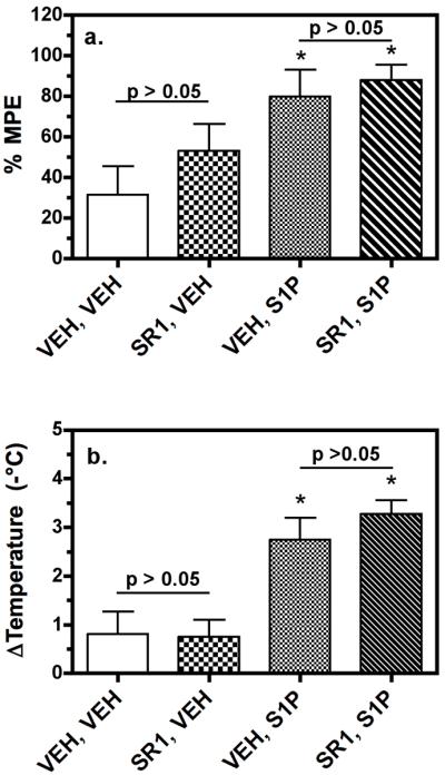
Sphingosine-1-phosphate (S1P) is a ubiquitous, lipophilic cellular mediator that acts in part by activation of G-protein-coupled receptor. Modulation of S1P signaling is an emerging pharmacotherapeutic target for immunomodulatory drugs. Although multiple S1P receptor types exist in the CNS, little is known about their function. Here, we report that S1P stimulated G-protein activity in the CNS, and results from [(35)S]GTPgammaS autoradiography using the S1P(1)-selective agonist SEW2871 and the S1P(1/3)-selective antagonist VPC44116 show that in several regions a majority of this activity is mediated by S1P(1) receptors. S1P receptor activation inhibited glutamatergic neurotransmission as determined by electrophysiological recordings in cortical neurons in vitro, and this effect was mimicked by SEW2871 and inhibited by VPC44116. Moreover, central administration of S1P produced in vivo effects resembling the actions of cannabinoids, including thermal antinociception, hypothermia, catalepsy and hypolocomotion, but these actions were independent of CB(1) receptors. At least one of the central effects of S1P, thermal antinociception, is also at least partly S1P(1) receptor mediated because it was produced by SEW2871 and attenuated by VPC44116. These results indicate that CNS S1P receptors are part of a physiologically relevant and widespread neuromodulatory system, and that the S1P(1) receptor contributes to S1P-mediated antinociception.


Figures

Figure 1
Figure 1
Representative autoradiograms and densitometric analysis reveal region-specific S1P-and SEW2871-stimulated G-protein activity in mouse brain. a.) Autoradiograms show basal binding in the absence of agonist (left column) and stimulation of [35S]GTPγS binding by 60 μM S1P (center column) or 80 μM SEW2871 (S1P1-selective agonist, right column). b.) Densitometric analysis of S1P-and SEW2871-stimulated [35S]GTPγS binding in the brain reveal regional differences in the magnitude of G-protein activation. Data are expressed as net agonist-stimulated activity ± SEM. **, SEW2871 was p < 0.01 different from S1P by 2-way ANOVA (drug x region) with post-hoc Bonferroni test (n = 10-12). c.) SEW2871-stimulated [35S]GTPγS binding, expressed as a percent of S1P-stimulated activity in the brain, varies among brain regions, suggesting that S1P1 receptors contribute varying proportions of the total G-protein activation seen with S1P in different brain regions. Regions with different letter designations are p < 0.05 different from each other by ANOVA with post-hoc Newman-Keuls test (n = 10-12). Abbreviations: Amyg; amygdala, Cblm; cerebellum, cc; corpus callosum, CCtx; cingulated cortex, CPu; caudate-putamen, Hip; hippocampus, Hyp; hypothalamus, PAG; periaqueductal gray, Thal; thalamus.
Figure 2
Figure 2
Net S1P- and SEW2871-stimulated G-protein activity in cerebellum and cingulate cortex is inhibited by the S1P1/3 antagonist VPC44116. Sections at the level of the a.) cerebellum or b.) cingulate cortex were incubated with vehicle (basal), S1P (10 μM) or SEW2871 (20 μM) in the presence and absence of the S1P1/3 antagonist VPC44116 (50 μM). VPC44116 blocked the stimulation of [35S]GTPγS binding by S1P or SEW2871, but did not affect [35S]GTPγS binding by itself. [35S]GTPγS binding is expressed as mean net stimulate [35S]GTPγS binding ± SEM. The p values shown were obtained by ANOVA with post-hoc Newman-Keuls test (n = 6-7).
Figure 3
Figure 3
S1P decreases sEPSC frequency and amplitude in cortical pyramidal neurons. a.) Whole-cell recording of sEPSCs (downward deflections) from an individual cortical pyramidal neuron. Application of 1 μM S1P (black bar) reduced sEPSC frequency and amplitude. b.) sEPSC parameters for each condition were normalized to the pre-drug value in each neuron. 1 μM S1P decreased mean sEPSC frequency by 27.3 ± 9.5% (n=11, p < 0.04 vs. control, ANOVA) and sEPSC frequency returned to 90.1 ± 9.5% of control frequency after removal of S1P. c.) 1 μM S1P decreased mean sEPSC amplitude by 12.7 ± 2.7% (n=10, p < 0.01 vs. control) and sEPSC amplitude returned to 94.7 ± 2.6% of control values after wash. d.) 1 μM S1P decreased mean sEPSC charge transfer by 42.0 ± 5.3% (n=10, p < 0.001 vs. control) and sEPSC charge transfer returned to 90.8 ± 10.8% of control values after wash. Statistical significance was determined by ANOVA with post-hoc Newman-Keuls test.
Figure 4
Figure 4
S1P-induced suppression of sEPSC activity is attenuated by the S1P1/3 receptor antagonist, VPC44116. a.) Whole-cell recording of sEPSCs from an individual cortical pyramidal neuron during the application of 1 μM S1P (black bar) and the subsequent addition of 5 μM VPC44116 (striped bar). The addition of VPC44116 partially attenuated the decrease in sEPSC frequency and amplitude induced by S1P. b.) sEPSC parameters for each condition were normalized to the pre-drug value in each neuron. 1 μM S1P decreased mean sEPSC frequency by 57.1± 16.7% (n=5, p < 0.03 vs. control) and the addition of 5 μM VPC44116 restored sEPSC frequency to 69.0 ± 21.6% of control frequency. c.) 1 μM S1P decreased mean median sEPSC amplitude by 25.0 ± 7.5% (n=5, p > 0.05 vs. control) and the addition of 5 μM VPC44116 restored sEPSC amplitude to 96.7 ± 15.8% of control amplitude. d.) 1 μM S1P decreased mean sEPSC charge transfer by 54.4 ± 9.1% (n=5, * p < 0.002 vs. control) and the addition of 5 μM VPC44116 restored sEPSC frequency to 87.3 ± 9.4% of control frequency (** p < 0.01 vs. S1P, p >0.05 vs. control). Statistical significance was determined by ANOVA with post-hoc Newman-Keuls test.
Figure 5
Figure 5
The S1P1 receptor agonist, SEW2871, decreases sEPSC activity. a.) Whole-cell recording of sEPSCs from an individual cortical pyramidal neuron during the application of 20 μM SEW2871 (black bar) and the subsequent addition of 5 μM VPC44116 (striped bar).b.) 20 μM SEW2871 decreased mean sEPSC charge transfer by 43.8 ± 8.2 % (n=8, * p < 0.05 vs. control by) and the administration of 5 μM VPC44116 in the continued presence of SEW2871 restored sEPSC charge transfer to 85.5 ± 10.1% of control values (n=8, ** p<0.05 vs. SEW2871 only, p> 0.05 vs. control). Statistical significance was determined by ANOVA with post-hoc Newman-Keuls test.
Figure 6
Figure 6
S1P produces antinociception to thermal stimulus of the tail in mice via S1P1 receptors. Mice were injected i.c.v with S1P with or without VPC44116. a. Dose-response curve for S1P-mediated antinociception in the tail-flick assay. S1P was injected i.c.v. and tail-flick latencies were obtained 20 min after injection. S1P produced dose-dependent antinociception. Data are mean %MPE values ± SEM. b. Antinociception expressed as mean %MPE ± SEM, determined following i.c.v. injection of S1P (25 μg) or the S1P1-selective agonist SEW2871 (SEW, 50 μg) with or without prior injection (i.c.v.) of vehicle or the S1P1/3 antagonist VPC44116 (25 μg). VPC44116 blocked the antinociceptive action of S1P or SEW2871, but administration of VPC44116 alone had no effect. Statistical significance of the data was determined by ANOVA with post-hoc Newman-Keuls test (n = 6).
Figure 7
Figure 7
S1P administered i.c.v. produces antinociception (a), hypothermia (b), catalepsy (c) and locomotor inhibition (d) in mice. Mice were injected (i.c.v.) with either vehicle or VPC44116 (25 μg) prior to injection (i.c.v.) with vehicle or S1P (25 μg), and tested following the final injection as described in Methods. S1P alone produced significant effects in all four tests. *, **, p < 0.05, 0.01 different from the vehicle/vehicle-treated group by ANOVA with post-hoc Dunnett's test (n = 6). VPC44116 significantly inhibited S1P-mediated antinociception (a.) as determined by ANOVA with post-hoc Newman-Keuls test (n = 6). VPC44116 alone produced catalepsy (d.) and partially inhibited S1P-induced locomotor inhibition (c.).
Figure 8
Figure 8
S1P-mediated antinociception and hypothermia does not require cannabinoid CB1 receptor activation. Mice were injected (i.p.) with 3mg/kg of the CB1-selective antagonist rimonabant (SR141716A) prior to injection (i.c.v.) of S1P (25 μg), and tested following the final injection as described in Methods. S1P produced significant antinociception (a.) and hypothermia (b.). *, **, p < 0.05, 0.01 different from the vehicle/vehicle-treated group by ANOVA with post-hoc Dunnett's test (n = 6). Rimonabant did not inhibit S1P-induced antinociception (a.) or hypothermia (b.) and rimonabant alone had no significant effects on either measure (a., b.), as determined by ANOVA with post-hoc Newman-Keuls test (n = 6).

References

    1. Beer MS, Stanton JA, Salim K, Rigby M, Heavens RP, Smith D, McAllister G. EDG receptors as a therapeutic target in the nervous system. Ann. N. Y. Acad. Sci. 2000;905:118–131. - PubMed
    1. Bliss CI. Statistics in Biology. McGraw-Hill; New York: 1967.
    1. Blondeau N, Lai Y, Tyndall S, Popolo M, Topalkara K, Pru JK, Zhang L, Kim H, Liao JK, Ding K, Waeber C. Distribution of sphingosine kinase activity and mRNA in rodent brain. J. Neurochem. 2007;103:509–517. - PMC - PubMed
    1. Brinkmann V. Sphingosine 1-phosphate receptors in health and disease: Mechanistic insights from gene deletion studies and reverse pharmacology. Pharmacol. Ther. 2007;115:84–105. - PubMed
    1. Bryan L, Kordula T, Spiegel S, Milstien S. Regulation and functions of sphingosine kinases in the brain. Biochim. Biophys. Acta. 2008 - PMC - PubMed

Publication types

MeSH terms