

Functional expression of the thyrotropin receptor in C cells: new insights into their involvement in the hypothalamic-pituitary-thyroid axis
- PMID: 19493188
- PMCID: PMC2740962
- DOI: 10.1111/j.1469-7580.2009.01095.x
Functional expression of the thyrotropin receptor in C cells: new insights into their involvement in the hypothalamic-pituitary-thyroid axis
Abstract
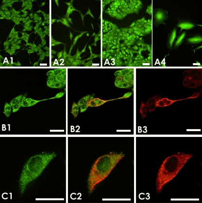
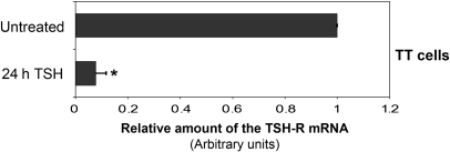
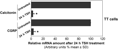
Thyroid C cells, or parafollicular cells, are mainly known for producing calcitonin, a hormone involved in calcium homeostasis with hypocalcemic and hypophosphatemic effects. Classically, the main endocrine activity of this cell population has been believed to be restricted to its roles in serum calcium and bone metabolism. Nonetheless, in the last few years evidence has been accumulating in the literature with regard to local regulatory peptides secreted by C cells, such as somatostatin, ghrelin, thyrotropin releasing hormone or the recently described cocaine- and amphetamine-related transcript, which could modify thyroid function. As thyrotropin is the main hormone controlling the hypothalamic-pituitary-thyroid axis and, accordingly, thyroid function, we have examined the functional expression of the thyrotropin receptor in C-cell lines and in thyroid tissues. We have found that rat and human C-cell lines express the thyrotropin receptor at both mRNA and protein levels. Furthermore, incubation of C cells with thyrotropin resulted in a 10-fold inhibition of thyrotropin-receptor expression, and a concomitant decrease of the steady-state mRNA levels for calcitonin and calcitonin gene-related peptide determined by quantitative real-time PCR was found. Finally, thyrotropin receptor expression by C cells was confirmed at protein level in both normal and pathological thyroid tissues by immunohistochemistry and immunofluorescence. These results confirm that C cells, under regulation by thyrotropin, are involved in the hypothalamic-pituitary-thyroid axis and suggest a putative role in local fine-tuning of follicular cell activity.
Figures


Similar articles
-
Expression of hypothalamic-pituitary-thyroid axis related genes in the human skin.J Invest Dermatol. 2002 Dec;119(6):1449-55. doi: 10.1046/j.1523-1747.2002.19617.x. J Invest Dermatol. 2002. PMID: 12485453 Free PMC article.
-
Thyroid-stimulating hormone, a novel, locally produced modulator of human epidermal functions, is regulated by thyrotropin-releasing hormone and thyroid hormones.Endocrinology. 2010 Apr;151(4):1633-42. doi: 10.1210/en.2009-0306. Epub 2010 Feb 22. Endocrinology. 2010. PMID: 20176727
-
Correction and Republication: Effect of Di-(2-ethylhexyl) phthalate on the hypothalamus-pituitary-thyroid axis in adolescent rat.Endocr J. 2018 Mar 28;65(3):261-268. doi: 10.1507/endocrj.EJ17-0272. Epub 2017 Dec 9. Endocr J. 2018. Corrected and republished in: Endocr J. 2022;69(2):217-224. doi: 10.1507/endocrj.EJ17-0272r. PMID: 29225205 Corrected and republished.
-
Hypothalamus-Pituitary-Thyroid Axis.Compr Physiol. 2016 Jun 13;6(3):1387-428. doi: 10.1002/cphy.c150027. Compr Physiol. 2016. PMID: 27347897 Review.
-
Syndromes of hormone resistance in the hypothalamic-pituitary-thyroid axis.Best Pract Res Clin Endocrinol Metab. 2006 Dec;20(4):529-46. doi: 10.1016/j.beem.2006.11.001. Best Pract Res Clin Endocrinol Metab. 2006. PMID: 17161330 Review.
Cited by
-
Medullary Thyroid Carcinoma: Recent Advances Including MicroRNA Expression.Endocr Pathol. 2016 Dec;27(4):312-324. doi: 10.1007/s12022-016-9449-0. Endocr Pathol. 2016. PMID: 27539727 Review.
-
Deficiency of 14-3-3ε and 14-3-3ζ by the Wnt1 promoter-driven Cre recombinase results in pigmentation defects.BMC Res Notes. 2016 Mar 22;9:180. doi: 10.1186/s13104-016-1980-z. BMC Res Notes. 2016. PMID: 27001213 Free PMC article.
-
Olfactory marker protein expression is an indicator of olfactory receptor-associated events in non-olfactory tissues.PLoS One. 2015 Jan 30;10(1):e0116097. doi: 10.1371/journal.pone.0116097. eCollection 2015. PLoS One. 2015. PMID: 25635859 Free PMC article.
-
Thyroid regeneration: characterization of clear cells after partial thyroidectomy.Endocrinology. 2012 May;153(5):2514-25. doi: 10.1210/en.2011-1365. Epub 2012 Mar 27. Endocrinology. 2012. PMID: 22454152 Free PMC article.
-
Subclinical Lipopolysaccharide from Salmonella Enteritidis Induces Dysregulation of Bioactive Substances from Selected Brain Sections and Glands of Neuroendocrine Axes.Toxins (Basel). 2019 Feb 2;11(2):91. doi: 10.3390/toxins11020091. Toxins (Basel). 2019. PMID: 30717384 Free PMC article.
References
-
- Abe E, Marians RC, Yu W, et al. TSH is a negative regulator of skeletal remodeling. Cell. 2003;115:151–162. - PubMed
-
- Ahren B. Effects of calcitonin, katacalcin, and calcitonin gene-related peptide on basal and TSH-stimulated thyroid hormone secretion in the mouse. Acta Physiol Scand. 1989;135:133–137. - PubMed
-
- Ahren B. Regulatory peptides in the thyroid gland – a review on their localization and function. Acta Endocrinol (Copenh) 1991;124:225–232. - PubMed
-
- Ain KB, Taylor KD, Tofiq S, Venkataraman G. Somatostatin receptor subtype expression in human thyroid and thyroid carcinoma cell lines. J Clin Endocrinol Metab. 1997;82:1857–1862. - PubMed
-
- Albores-Saavedra J, Monforte H, Nadji M, Morales AR. C-cell hyperplasia in thyroid tissue adjacent to follicular cell tumors. Hum Pathol. 1988;19:795–799. - PubMed
Publication types
MeSH terms
Substances
LinkOut - more resources
Full Text Sources
Research Materials
