Review
. 2009 Aug;22(4):400-19.
doi: 10.1111/j.1755-148X.2009.00585.x. Epub 2009 May 28.

The PTEN-AKT3 signaling cascade as a therapeutic target in melanoma

Affiliations
Review

The PTEN-AKT3 signaling cascade as a therapeutic target in melanoma

Subbarao V Madhunapantula et al. Pigment Cell Melanoma Res. 2009 Aug.

Abstract

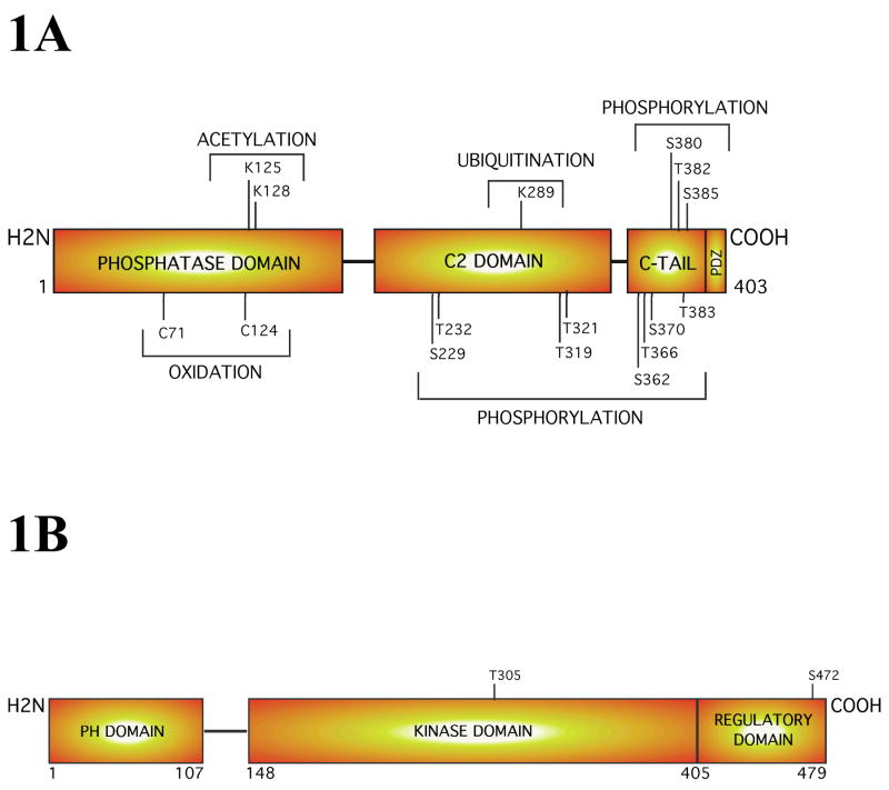
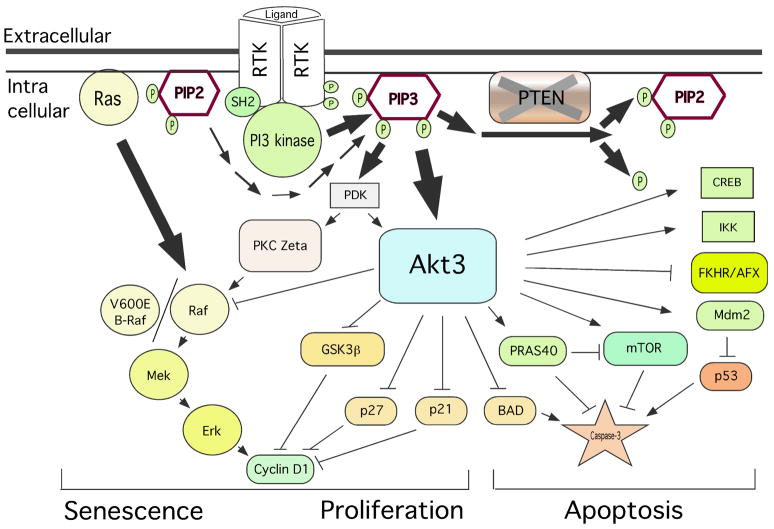
Melanocytes undergo extensive genetic changes during transformation into aggressive melanomas. These changes deregulate genes whose aberrant activity promotes the development of this disease. The phosphoinositide-3-kinase (PI3K) and mitogen-activated protein (MAP) kinase pathways are two key signaling cascades that have been found to play prominent roles in melanoma development. These pathways relay extra-cellular signals via an ordered series of consecutive phosphorylation events from cell surface throughout the cytoplasm and nucleus regulating diverse cellular processes including proliferation, survival, invasion and angiogenesis. It is generally accepted that therapeutic agents would need to target these two pathways to be an effective therapy for the long-term treatment of advanced-stage melanoma patients. This review provides an overview of the PI3 kinase pathway focusing specifically on two members of the pathway, called PTEN and Akt3, which play important roles in melanoma development. Mechanisms leading to deregulation of these two proteins and therapeutic implications of targeting this signaling cascade to treat melanoma are detailed in this review.


Figures

Figure 1
Figure 1. Structure of the PTEN and Akt3 proteins
1A PTEN, a tumor suppressor protein and lipid phosphatase, is a 55 kDa enzyme containing an N-terminal phosphatase domain, a central C2 domain, and a C-terminal tail. Each domain has sites for post-translational modifications that could involve: acetylation, oxidation, ubiquitination, or phosphorylation, which have potential to regulate PTEN activity. 1B. Structural analysis of Akt3 reveals that this oncogenic survival kinase increasingly active during melanoma progression consists of an N-terminal PH domain, a central kinase domain and a C-terminal regulatory motif. Complete activation of this kinase occurs only when the threonine 308 (T308) and serine 472 (S472) gets phosphorylated by PDK1 and a PDK2.
Figure 2
Figure 2. PI3K and MAPK signaling interactions in melanoma
PI3 and MAP kinase signaling cascades regulate cell senescence, proliferation and apoptosis. Whereas the Ras/Raf/Mek/Erk signaling arm is primarily involved in controlling proliferation, it also induce cell senescence in benign nevi to inhibit further progression into melanomas. The survival kinase Akt3 inhibits apoptosis and induces proliferation by phosphorylation of substrate proteins. Recently, Akt3 has been shown to inhibit V600EB-Raf activity by phosphorylation thereby promoting early melanoma development.
Figure 3
Figure 3. PTEN regulates apoptosis by inhibiting Akt activity in melanomas
3A PTEN expression decreases pAkt levels in melanoma ccells lacking functional PTEN. UACC 903 melanoma cells lacking functional PTEN protein were infected with increasing amounts (7.8, 15.6, 31.3, 62.5, 125 μL) of adeno-associated viral constructs containing Wt PTEN, HA tagged Wt PTEN and a catalytically inactive G129R PTEN mutation. Three days later, cell lysates were collected and proteins analyzed by Western blotting. Levels of pAkt and total Akt were quantitated by densitometry and the pAkt/totalAkt ratio represented against amount of viral supernatant used for infection. Data show that pAkt expression decreases with increasing virus amount indicating PTEN reduces pAkt in melanomas (Stahl et al., 2003). 3B. PTEN triggers apoptosis in melanomas. Levels of cleaved caspase-3, an indicator of apoptosis, were measured using Western blotting following expression of viral introduced PTEN into UACC 903 melanoma cells lacking functional PTEN protein. Compared to functionally inactive G129R PTEN, wild type PTEN expression increased levels of cleaved caspase-3 indicating elevatedlevels of apoptosis. α-enolase served as control for protein loading (Stahl et al., 2003).
Figure 4
Figure 4. PTEN loss leads to preferential increase in Akt3 activity in melanomacytes and radial growth phase melananoma cells
Normal human melanocytes and a cell line derived from a radial growth phase of melanoma (WM35) that contain low pAkt levels were transfected with siRNA targeting PTEN and/or one of the isoforms of Akt. A significant decrease in pAkt was noticed only when PTEN and Akt3 were targeted together but not with either PTEN and Akt1 or PTEN and Akt2, indicating that Akt3 is activated preferentially following PTEN loss (Stahl et al., 2004).
Figure 5
Figure 5. Akt3 expression and activity increase during melanoma development
5A Expression of pAkt in nevi, dysplastic nevi, primary and metastatic melanoma lesions was quantitated from histological sections and intensity scored using immunohistochemical staining. Compared to common nevi that had only weak-to-moderate pAkt staining, increasing percentages of dysplastic nevi, primary and metastatic melanomas contained higher levels of Akt activity with 67% of advanced tumors having elevated activity (Stahl et al., 2004). 5B. Expression and activity of Akt isoforms (Akt1, Akt2 and Akt3) was measured in flash frozen melanoma patient tumor samples by Western blotting. Approximattely 60% of melanoma tumors expressed elevated levels of Akt3 protein compared to normal human melanocytes. Furthermore, ~43% of tumor had elevated Akt3 activity.
Figure 6
Figure 6. PTEN introduction or Akt3 inhibition induce apoptosis in melanomas
Photographs shows TUNEL-positive cells (an indicator of apoptosis) in tumor xenografts following PTEN expression (6A) or siRNA-mediated reduction of Akt3 protein levels (6B). UACC 903 cells or cells expressing chromosomal PTEN or cells nucleofected with siAkt3 versus siAkt2 (1 × 10⁶) were injected into nude mice and 4 days later processed for TUNEL staining. Data shows that either PTEN expression or Akt3 protein knockdown increased apopotosis compared to controls in which PTEN activity had been lost (revertant) or cells with reduced Akt2 protein levels (Stahl et al., 2004).
Figure 7
Figure 7. Akt3 activity promotes melanoma development by inhibiting V600EB-Raf
Akt3 decreases V600EB-Raf activity by phosphorylating the protein thereby decreasing its activity and that of the downstream signaling cascade to levels that promote rather than inhibit melanocyte proliferation, which aids melanoma development.
Figure 8
Figure 8. Knockdown of PRAS40, a substrate of Akt3, inhibits melanoma tumor growth
SiRNA-mediated reduction of PRAS40 protein levels, a direct substrate of Akt, or upstream Akt3 inhibits tumor development in xenografted melanoma tumors. UACC 903 cells were transfected with siRNAs targeting PRAS40 or Akt3, and 1.5 days later cells were injected into nude mice. Developing tumor sizes were measured on alternate days. Compared to buffer, a scrambled siRNA or a siRNA targeting C-Raf, a 50–60% decrease in tumor volume observed following PRAS40 or Akt3 protein knockdown. Inset shows reduction of PRAS40 protein levels in tumors removed from mice at days 7.5 or 9.5 (Madhunapantula et al., 2007).
Figure 9
Figure 9
ISC-4 inhibits melanoma tumor development by targeting Akt3 signaling. 10A. Structures of 4 and 6 carbon containing isothiocyanates (PBITC and PHITC) and isoselenocyanates (ISC-4 and ISC6). 9B. Isoselenocyanates (ISC-4 and ISC-6) were injected intra peritoneally into mice bearing established 50–60 mm3 sub-cutaneous melanoma tumors. Compared to DMSO vehicle controls or PBITC and PHITC, isoselenocyanates decreased tumor development by ~50% (Sharma et al., 2009). 9B. ISC-4 reduced Akt3 signaling in melanoma tumors. Decreased pAkt3 and downstream pPRAS40 were observed in tumors treated with isoselenocyanates indicating the compound decreased Akt3 signaling in tumors (Sharma et al., 2009).
Figure 10
Figure 10. Targeting Akt3 and V600EB-Raf synergistically inhibits melanoma tumor development
SiRNAs targeting Akt3 (100 picomoles) and V600EB-Raf (12.5 picomoles) were introduced into UACC 903 melanoma cells alone or in combination, which were injected subcutaneously in to mice and tumor development measured on alternate days upto days 17.5. Compared to controls (buffer, siScrambled or siC-Raf) an ~60% reduction in tumor development was observed targeting Akt3 or V600EB-Raf alone. A much more dramatic reduction (~80%) was when these proteins were targeted together. Inset shows decreased Akt3 and V600EB-Raf protein expression levels in melanoma tumors at days 9.5 (Cheung et al., 2008).

Similar articles

Cited by

References

    1. Al-Khouri AM, Ma Y, Togo SH, Williams S, Mustelin T. Cooperative phosphorylation of the tumor suppressor phosphatase and tensin homologue (PTEN) by casein kinases and glycogen synthase kinase 3beta. J Biol Chem. 2005;280:35195–35202. - PubMed
    1. Alessi DR, Andjelkovic M, Caudwell B, Cron P, Morrice N, Cohen P, Hemmings BA. Mechanism of activation of protein kinase B by insulin and IGF-1. Embo J. 1996;15:6541–6551. - PMC - PubMed
    1. Alessi DR, Cuenda A, Cohen P, Dudley DT, Saltiel AR. PD 098059 is a specific inhibitor of the activation of mitogen-activated protein kinase kinase in vitro and in vivo. J Biol Chem. 1995;270:27489–27494. - PubMed
    1. Andjelkovic M, Alessi DR, Meier R, Fernandez A, Lamb NJ, Frech M, Cron P, Cohen P, Lucocq JM, Hemmings BA. Role of translocation in the activation and function of protein kinase B. J Biol Chem. 1997;272:31515–31524. - PubMed
    1. Andjelkovic M, Jakubowicz T, Cron P, Ming XF, Han JW, Hemmings BA. Activation and phosphorylation of a pleckstrin homology domain containing protein kinase (RAC-PK/PKB) promoted by serum and protein phosphatase inhibitors. Proc Natl Acad Sci U S A. 1996;93:5699–5704. - PMC - PubMed

Publication types

MeSH terms