. 2009 Oct;22(5):623-34.
doi: 10.1111/j.1755-148X.2009.00582.x. Epub 2009 May 26.

Agouti protein, mahogunin, and attractin in pheomelanogenesis and melanoblast-like alteration of melanocytes: a cAMP-independent pathway

Affiliations
Free PMC article

Agouti protein, mahogunin, and attractin in pheomelanogenesis and melanoblast-like alteration of melanocytes: a cAMP-independent pathway

Tokimasa Hida et al. Pigment Cell Melanoma Res. 2009 Oct.
Free PMC article

Abstract

Melanocortin-1 receptor (MC1R) and its ligands, alpha-melanocyte stimulating hormone (alphaMSH) and agouti signaling protein (ASIP), regulate switching between eumelanin and pheomelanin synthesis in melanocytes. Here we investigated biological effects and signaling pathways of ASIP. Melan-a non agouti (a/a) mouse melanocytes produce mainly eumelanin, but ASIP combined with phenylthiourea and extra cysteine could induce over 200-fold increases in the pheomelanin to eumelanin ratio, and a tan-yellow color in pelletted cells. Moreover, ASIP-treated cells showed reduced proliferation and a melanoblast-like appearance, seen also in melanocyte lines from yellow (A(y)/a and Mc1r(e)/ Mc1r(e)) mice. However ASIP-YY, a C-terminal fragment of ASIP, induced neither biological nor pigmentary changes. As, like ASIP, ASIP-YY inhibited the cAMP rise induced by alphaMSH analog NDP-MSH, and reduced cAMP level without added MSH, the morphological changes and depigmentation seemed independent of cAMP signaling. Melanocytes genetically null for ASIP mediators attractin or mahogunin (Atrn(mg-3J/mg-3J) or Mgrn1(md-nc/md-nc)) also responded to both ASIP and ASIP-YY in cAMP level, while only ASIP altered their proliferation and (in part) shape. Thus, ASIP-MC1R signaling includes a cAMP-independent pathway through attractin and mahogunin, while the known cAMP-dependent component requires neither attractin nor mahogunin.


Figures
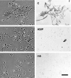
Figure 1
Figure 1
Morphological and pigmentary changes induced in melan-a cells by agouti signaling protein (ASIP). Paired phase contrast (left) and bright-field (right, to show eumelanin) images of melan-a cells grown in normal growth medium (C, control); or in the same medium with 10 nM ASIP added for the last 5 days (ASIP). No phenylthiourea (PTU) or extra cysteine was present (see text). (mb): normal murine melanoblasts in a primary neonatal epidermal melanocyte culture, 7 days after explantation, showing similarity to melan-a cells grown with ASIP. Bar = 100 μm.
Figure 2
Figure 2
Marked increases in pheomelanin/eumelanin ratio and tan coloration by agouti signaling protein (ASIP), cysteine and phenylthiourea (PTU). Cells were subcultured as needed to prevent confluency. (A) Eumelanin and pheomelanin content of melan-a cells after 14 days’ growth with 300 μM PTU followed by 14 days’ growth in standard medium [containing 12-O-tetradecanoyl phorbol-13-acetate (TPA)] with the indicated additives. Data show mean ± SEM of three different samples. (ASIP): 10 nM ASIP; (Cys): 2 mM cysteine plus 100 μM mercaptoethanol; (PTU): 100 μM PTU. (B) Pellets of cells after 14 days’ growth with 500 μM PTU followed by 5 days’ growth with the indicated additives. (A/C): 10 nM ASIP, 2 mM cysteine and 100 μM mercaptoethanol; (TPA): 200 nM TPA; (PTU): PTU at 500 μM for 2 days and 100 μM for 3 days. Significances of selected differences by Student’s t-test (two-tailed) are shown: *P < 0.05; **P < 0.01; ***P < 0.001, (NS): P > 0.05, not significant.
Figure 3
Figure 3
Melanoblast-like appearance of melan-Ay and melan-e cells in the absence of CT. Paired phase-contrast (left) and bright-field (right) images of melan-Ay (Ay) and melan-e (e) cells grown with 20 pM CT (CT) or for several passages without CT (0). Some residual eumelanin is seen in some cells of both genotypes without CT. For reference melan-a cells (a) grown with 20 pM CT are also shown; there is little difference from melan-a cells without CT (Figure 1). Bar = 100 μm.
Figure 5
Figure 5
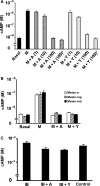
(A) Inhibition of NDP-MSH-induced cAMP rise by both agouti signaling protein (ASIP) and ASIP-YY in melan-a cells. Intracellular cAMP per well was measured in cultures that had been plated and allowed to attach overnight, after stimulation for 30 min with 30 pM NDP-MSH (M), with or without ASIP (A) or ASIP-YY (Y). Figures in parentheses are concentration of ASIP or ASIP-YY in nM. *RPMI1640 medium with 10% FBS was incubated with ASIP or ASIP-YY at 37°C overnight and was used as stimulating buffer, followed by application of NDP-MSH. Shown are mean ± SEM of triplicate wells. (B) Inhibition of NDP-MSH-induced cAMP rise by both ASIP and ASIP-YY in melan-mg and melan-md cells. Assay as above, using 100 nM ASIP (A) or 100 nM ASIP-YY (Y). The reason for the lower apparent stimulation of melan-a cells by NDP-MSH in (B) than in (A) is unknown; it may reflect change in the assay reagents with time. However the relative responses of different genotypes are clear. (C) Reduction of basal cAMP by both ASIP and ASIP-YY. In this experiment only, to increase assay sensitivity, melan-a cells were plated at a higher number of 6 × 10⁴ cells/well, preincubation with isobutylmethylxanthine (IBMX) was for 5 h at 1 μM, and incubation with the stated additives was for 5 h. IBMX alone (1 μM, a low concentration) had no significant effect, whereas both ASIP (A) and ASIP-YY (Y) at 10 nM gave significant reductions in cAMP levels compared with 1 μM IBMX alone (P = 0.013, P = 0.003 respectively). Similar effects were seen under slightly varied conditions (not shown).
Figure 4
Figure 4
Melanoblast-like shape-change, depigmentation and growth inhibition of melan-a cells by agouti signaling protein (ASIP) but not ASIP-YY. (A) phase-contrast images of melan-a cells grown for 5 days in growth medium (0), with 10 nM ASIP (ASIP) or with 100 nM ASIP-YY (ASIP-YY). Bar = 100 μm. (B) Total melanin content of melan-a cells grown in growth medium only, or with ASIP or ASIP-YY for 14 days. Mean and SEM of triplicate dishes. (C) Cell doubling times of melan-a cells subcultured with no addition, ASIP or ASIP-YY for 30 days. Shown are mean ± SEM of triplicates.
Figure 6
Figure 6
Melanoblastic shape-change and growth inhibition of melan-mg and melan-md cells by agouti signaling protein (ASIP) but not ASIP-YY. Melan-mg cells (mg) and melan-md cells (md) were incubated for 5 days in growth medium alone (0), with 10 nM ASIP (ASIP) or with 10 nM ASIP-YY (YY). Bar = 100 μm. No Cys or phenylthiourea (PTU) present. Cell doubling times of melan-mg cells (B) and melan-md cells (C) were measured in the presence of 10 nM ASIP or 10 nM ASIP-YY for 30 days. Shown are mean ± SEM of triplicate cultures. Retardation of proliferation by ASIP was highly significant for both genotypes by Student’s t-test (P < 0.001), while ASIP-YY had no significant effect.
Figure 7
Figure 7
Model to account for responsiveness to cAMP level in activation but not inactivation of eumelanin synthesis. See Discussion for main explanation. (A-YY) ASIP-YY, (Mc) melanocyte, (Mb) melanoblast, (Ub) ubiquitinylation. Melanocortin-1 receptor (MC1R) is shown with some basal activity (left, lost in e mutant), to explain why POMC null, a/a mice are black, and related data (see text). (This might alternatively be due to BD103 or some unknown signal). Agouti signaling protein (ASIP) would not be required for pheomelanogenesis unless Y has been switched on by the MC1R/cAMP pathway, hence inactive MC1R would give a yellow color even with deficient MGRN1, as seen in e/e, md/md mice.

Similar articles

Cited by

References

    1. Abdel-Malek ZA, Scott MC, Furumura N, Lamoreux ML, Ollmann M, Barsh GS, Hearing VJ. The melanocortin 1 receptor is the principal mediator of the effects of agouti signaling protein on mammalian melanocytes. J. Cell Sci. 2001;114:1019–1024. - PubMed
    1. Aberdam E, Bertolotto C, Sviderskaya EV, de Thillot V, Hemesath TJ, Fisher DE, Bennett DC, Ortonne JP, Ballotti R. Involvement of microphthalmia in the inhibition of melanocyte lineage differentiation and of melanogenesis by Agouti signal protein. J. Biol. Chem. 1998;273:19560–19565. - PubMed
    1. Barsh GS. Regulation of pigment type switching by agouti, melanocortin signaling, attractin and mahoganoid. In: Nordlund JJ, Boissy RE, Hearing VJ, King RA, Oetting WS, Ortonne JP, editors. The Pigmentary System. 2nd edn. Massachusetts: Blackwell Publishing; 2006. pp. 395–409.
    1. Bennett DC. Mechanisms of differentiation in melanoma cells and melanocytes. Environ. Health Perspect. 1989;80:49–60. - PMC - PubMed
    1. Bennett DC, Lamoreux ML. The color loci of mice– a genetic century. Pigment Cell Res. 2003;16:333–344. - PubMed

Publication types

MeSH terms