. 2009 Aug;83(16):7970-81.
doi: 10.1128/JVI.00521-09. Epub 2009 Jun 3.

Determinants of hepatitis C virus p7 ion channel function and drug sensitivity identified in vitro

Affiliations

Determinants of hepatitis C virus p7 ion channel function and drug sensitivity identified in vitro

Corine StGelais et al. J Virol. 2009 Aug.

Abstract

Hepatitis C virus (HCV) chronically infects 170 million individuals, causing severe liver disease. Although antiviral chemotherapy exists, the current regimen is ineffective in 50% of cases due to high levels of innate virus resistance. New, virus-specific therapies are forthcoming although their development has been slow and they are few in number, driving the search for new drug targets. The HCV p7 protein forms an ion channel in vitro and is critical for the secretion of infectious virus. p7 displays sensitivity to several classes of compounds, making it an attractive drug target. We recently demonstrated that p7 compound sensitivity varies according to viral genotype, yet little is known of the residues within p7 responsible for channel activity or drug interactions. Here, we have employed a liposome-based assay for p7 channel function to investigate the genetic basis for compound sensitivity. We demonstrate using chimeric p7 proteins that neither the two trans-membrane helices nor the p7 basic loop individually determines compound sensitivity. Using point mutation analysis, we identify amino acids important for channel function and demonstrate that null mutants exert a dominant negative effect over wild-type protein. We show that, of the three hydrophilic regions within the amino-terminal trans-membrane helix, only the conserved histidine at position 17 is important for genotype 1b p7 channel activity. Mutations predicted to play a structural role affect both channel function and oligomerization kinetics. Lastly, we identify a region at the p7 carboxy terminus which may act as a specific sensitivity determinant for the drug amantadine.


Figures
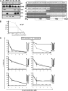
FIG. 1.
FIG. 1.
Drug sensitivity of chimeric J4/JFH-1 p7 proteins. Chimeric p7 proteins comprising J4 and JFH-1 amino and carboxy termini were expressed and purified from E. coli and assessed for their sensitivity to amantadine or rimantadine compared to the parental proteins. (A) Purified proteins were subjected to SDS-PAGE and Western analysis (left panel) using genotype-specific antibodies to J4 (no. 1055) or JFH-1 (no. 2717) carboxy termini or the JFH-1 amino terminus (no. 2715). Proteins were also probed with anti-FLAG monoclonal antibody and stained with Coomassie brilliant blue (CB). A schematic shows chimeric amino acid sequences for all four chimeras and parental proteins (right panel). White boxes, J4 sequence; black boxes, JFH-1 sequence. (B) A 50% inhibitory concentration curve for amantadine inhibition of J4 p7 in liposome CF release assays. (C) Liposome CF release assays for parental and chimeric proteins in the presence of increasing concentrations of amantadine or rimantadine. Activity is expressed as percent initial rate of untreated wild-type p7, measured by the ΔRFU/min for the first 5 min. The dotted line shows baseline dye release in solvent or liposome-only controls. Assays were performed for at least three separate batches of each protein with each condition in quadruplicate.
FIG. 2.
FIG. 2.
Expression, purification, and activities of mutated J4 p7 proteins. (A) Key p7 residues were selected for alanine substitution mutagenesis as described in the Results section (top panel). HPLC-purified cleaved protein (5 μg) was then subjected to SDS-PAGE and Western blot analysis using an anti-FLAG monoclonal antibody and the J4-specific antibody (no. 1055) (bottom panels). Proteins were also stained with Coomassie brilliant blue (CB). Lanes 1, wild type J4 FLAG-p7; lanes 2, K33A/R35A; lanes 3, S21A; lanes 4, C27A; lanes 5, F22/25/26A; lanes 6, G39A; lanes 7, P49A; lanes 8, L(50-55)A; lanes 9, H17A. TM1 and TM2, trans-membrane helixes 1 and 2. (B) Activities of alanine substitution mutants of J4 p7 were assessed in the liposome dye release assay in the presence (+Ama) or absence (no drug) of amantadine (1 μM). Activity is expressed as the percent initial rate of untreated wild-type p7, measured by the ΔRFU/min for the first 5 min. Assays were performed for at least three separate batches of each protein with each condition in quadruplicate. Asterisks indicate P values generated from a Student t test where applicable. Baseline dye release values in solvent (MeOH) and liposome-only (Lip) controls are shown. α, anti.
FIG. 3.
FIG. 3.
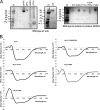
Oligomerization and folding of p7 null mutant proteins. (A) Mild detergents were tested for their ability to induce oligomerization of wild-type J4 FLAG-p7 during native PAGE. Both DHPC and DPC (300 mM) induced heptamer formation, whereas LMPG and LPPG did not (left panel). Wild-type protein was mixed with 50 μM liposomes (LIP) and analyzed by native PAGE (middle). Null-mutant p7 proteins were reconstituted in 300 mM DHPC, and oligomeric forms were resolved by native PAGE (right). (B) Wild-type and null-mutant proteins dissolved in methanol were assessed for alpha-helical content using CD. WT, wild type; M, molecular mass markers (kDa).
FIG. 4.
FIG. 4.
Membrane association and dominant negative effects of mutant J4 p7 proteins. (A) p7-null mutations were assessed for membrane association in the presence or absence of high pH (100 mM Na2CO3 [pH 11.4]). Anti-FLAG Western blots from fractionated discontinuous Ficoll gradients are shown from 1 (bottom) to 12 (top). Bottom panels show rhodamine fluorescence of gradient fractions with liposomes floating to the 10% Ficoll-aqueous interface (fraction 11). (B) Basic loop K33A/R35A and H17A mutant proteins were tested for their ability to exert a dominant negative effect over wild-type protein by mixing in increments of one-seventh of the total 5 μg of input compared to the corresponding amount of wild-type protein alone and testing resultant channel activity in the liposome dye release assay. Activity is expressed as the percent initial rate of untreated wild-type p7, measured by the ΔRFU/min for the first 5 min.
FIG. 5.
FIG. 5.
Effect of protein concentration on p7-null mutant dye release. Increasing amounts of null-mutant proteins were titrated into the dye release assay from 20 to 200% standard input levels. (A) Channel activity is expressed as the percent untreated wild-type (WT) protein initial rate under standard conditions (100% is equivalent to 5 μg in 100 μl, giving a final concentration of ∼5 μM). (B) Western blot analysis of wild-type, G39A, and P49A proteins taken following titration experiments showing higher-molecular-mass oligomeric forms of p7 in SDS micelles (arrows). One-tenth of the total amounts of protein used in the experiment described in panel A was loaded.
FIG. 6.
FIG. 6.
Decreased sensitivity of a polyleucine mutation L(50-55)A to low concentrations of amantadine but not rimantadine. Polyleucine mutant protein was subjected to 1 or 5 μM rimantadine (rim) or amantadine (ama), and channel activity was assessed in liposome dye release assays. The lower panel shows a comparison of amino acids in the carboxy terminus for J4, the L(50-55)A mutant, and JFH-1 1 proteins.
FIG. 7.
FIG. 7.
Molecular modeling of the J4 p7 ion channel complex. J4 p7 protomers were modeled with free energy minimization using the Maestro program and then manually docked into a symmetrical heptameric complex. The top panel shows top-down views and a side projection (two protomers removed) of channel complexes with the potential pH sensor, His 17, and possible gate, Tyr 31, highlighted. In addition, the positions of Pro 49 and Leu 50 to Leu 55(Poly-L) are indicated on the side projection to illustrate the interactions with adjacent protomers. Last, bottom views of the complex are shown, illustrating the apposed basic residues of the cytosolic loop, which may promote the hairpin conformation of each protomer.

Similar articles

Cited by

References

    1. Carrere-Kremer, S., C. Montpellier-Pala, L. Cocquerel, C. Wychowski, F. Penin, and J. Dubuisson. 2002. Subcellular localization and topology of the p7 polypeptide of hepatitis C virus. J. Virol. 763720-3730. - PMC - PubMed
    1. Chew, C. F., R. Vijayan, J. Chang, N. Zitzmann, and P. C. Biggin. 2009. Determination of pore-lining residues in the hepatitis C virus p7 protein. Biophys. J. 96L10-L12. - PMC - PubMed
    1. Choo, Q. L., G. Kuo, A. J. Weiner, L. R. Overby, D. W. Bradley, and M. Houghton. 1989. Isolation of a cDNA clone derived from a blood-borne non-A, non-B viral hepatitis genome. Science 244359-362. - PubMed
    1. Clarke, D., S. Griffin, L. Beales, C. S. Gelais, S. Burgess, M. Harris, and D. Rowlands. 2006. Evidence for the formation of a heptameric ion channel complex by the hepatitis C virus p7 protein in vitro. J. Biol. Chem. 28137057-37068. - PMC - PubMed
    1. Cocquerel, L., C. Wychowski, F. Minner, F. Penin, and J. Dubuisson. 2000. Charged residues in the transmembrane domains of hepatitis C virus glycoproteins play a major role in the processing, subcellular localization, and assembly of these envelope proteins. J. Virol. 743623-3633. - PMC - PubMed

Publication types

LinkOut - more resources