

Simian immunodeficiency virus SIVrcm, a unique CCR2-tropic virus, selectively depletes memory CD4+ T cells in pigtailed macaques through expanded coreceptor usage in vivo
- PMID: 19493994
- PMCID: PMC2715763
- DOI: 10.1128/JVI.00444-09
Simian immunodeficiency virus SIVrcm, a unique CCR2-tropic virus, selectively depletes memory CD4+ T cells in pigtailed macaques through expanded coreceptor usage in vivo
Abstract
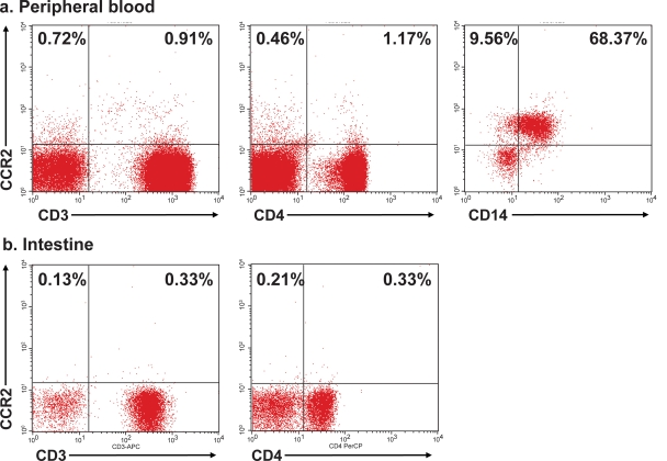
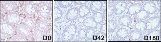
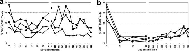
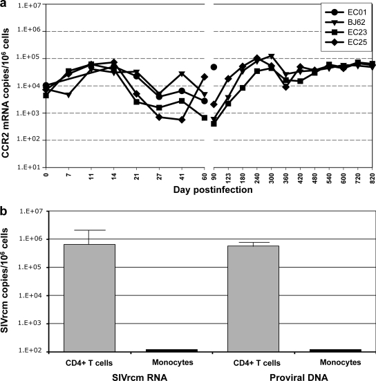
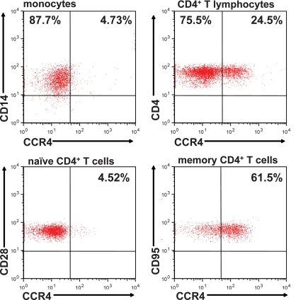
Simian immunodeficiency virus SIVrcm, which naturally infects red-capped mangabeys (RCMs), is the only SIV that uses CCR2 as its main coreceptor due to the high frequency of a CCR5 deletion in RCMs. We investigated the dynamics of SIVrcm infection to identify specific pathogenic mechanisms associated with this major difference in SIV biology. Four pigtailed macaques (PTMs) were infected with SIVrcm, and infection was monitored for over 2 years. The dynamics of in vivo SIVrcm replication in PTMs was similar to that of other pathogenic and nonpathogenic lymphotropic SIVs. Plasma viral loads (VLs) peaked at 10(7) to 10(9) SIVrcm RNA copies/ml by day 10 postinoculation (p.i.). A viral set point was established by day 42 p.i. at 10(3) to 10(5) SIVrcm RNA copies/ml and lasted up to day 180 p.i., when plasma VLs decreased below the threshold of detection, with blips of viral replication during the follow-up. Intestinal SIVrcm replication paralleled that of plasma VLs. Up to 80% of the CD4(+) T cells were depleted by day 28 p.i. in the gut. The most significant depletion (>90%) involved memory CD4(+) T cells. Partial CD4(+) T-cell restoration was observed in the intestine at later time points. Effector memory CD4(+) T cells were the least restored. SIVrcm strains isolated from acutely infected PTMs used CCR2 coreceptor, as reported, but expansion of coreceptor usage to CCR4 was also observed. Selective depletion of effector memory CD4(+) T cells is in contrast with predicted in vitro tropism of SIVrcm for macrophages and is probably due to expansion of coreceptor usage. Taken together, these findings emphasize the importance of understanding the selective forces driving viral adaptation to a new host.
Figures


Similar articles
-
Pathogenic features associated with increased virulence upon Simian immunodeficiency virus cross-species transmission from natural hosts.J Virol. 2014 Jun;88(12):6778-92. doi: 10.1128/JVI.03785-13. Epub 2014 Apr 2. J Virol. 2014. PMID: 24696477 Free PMC article.
-
V3 loop-determined coreceptor preference dictates the dynamics of CD4+-T-cell loss in simian-human immunodeficiency virus-infected macaques.J Virol. 2005 Oct;79(19):12296-303. doi: 10.1128/JVI.79.19.12296-12303.2005. J Virol. 2005. PMID: 16160156 Free PMC article.
-
Elite Control, Gut CD4 T Cell Sparing, and Enhanced Mucosal T Cell Responses in Macaca nemestrina Infected by a Simian Immunodeficiency Virus Lacking a gp41 Trafficking Motif.J Virol. 2015 Oct;89(20):10156-75. doi: 10.1128/JVI.01134-15. Epub 2015 Jul 29. J Virol. 2015. PMID: 26223646 Free PMC article.
-
The Hitchhiker Guide to CD4+ T-Cell Depletion in Lentiviral Infection. A Critical Review of the Dynamics of the CD4+ T Cells in SIV and HIV Infection.Front Immunol. 2021 Jul 21;12:695674. doi: 10.3389/fimmu.2021.695674. eCollection 2021. Front Immunol. 2021. PMID: 34367156 Free PMC article.
-
SIV Coreceptor Specificity in Natural and Non-Natural Host Infection: Implications for Cell Targeting and Differential Outcomes from Infection.Curr HIV Res. 2018;16(1):41-51. doi: 10.2174/1570162X15666171124121805. Curr HIV Res. 2018. PMID: 29173179 Review.
Cited by
-
Pathogenicity and mucosal transmissibility of the R5-tropic simian/human immunodeficiency virus SHIV(AD8) in rhesus macaques: implications for use in vaccine studies.J Virol. 2012 Aug;86(16):8516-26. doi: 10.1128/JVI.00644-12. Epub 2012 May 30. J Virol. 2012. PMID: 22647691 Free PMC article.
-
Nonhuman Primate Testing of the Impact of Different Regulatory T Cell Depletion Strategies on Reactivation and Clearance of Latent Simian Immunodeficiency Virus.J Virol. 2020 Sep 15;94(19):e00533-20. doi: 10.1128/JVI.00533-20. Print 2020 Sep 15. J Virol. 2020. PMID: 32669326 Free PMC article.
-
Walk on the wild side: SIV infection in African non-human primate hosts-from the field to the laboratory.Front Immunol. 2023 Jan 12;13:1060985. doi: 10.3389/fimmu.2022.1060985. eCollection 2022. Front Immunol. 2023. PMID: 36713371 Free PMC article. Review.
-
Cloning and analysis of sooty mangabey alternative coreceptors that support simian immunodeficiency virus SIVsmm entry independently of CCR5.J Virol. 2012 Jan;86(2):898-908. doi: 10.1128/JVI.06415-11. Epub 2011 Nov 16. J Virol. 2012. PMID: 22090107 Free PMC article.
-
Pathogenic features associated with increased virulence upon Simian immunodeficiency virus cross-species transmission from natural hosts.J Virol. 2014 Jun;88(12):6778-92. doi: 10.1128/JVI.03785-13. Epub 2014 Apr 2. J Virol. 2014. PMID: 24696477 Free PMC article.
References
-
- Agy, M. B., L. R. Frumkin, L. Corey, R. W. Coombs, S. M. Wolinsky, J. Koehler, W. R. Morton, and M. G. Katze. 1992. Infection of Macaca nemestrina by human immunodeficiency virus type-1. Science 257103-106. - PubMed
-
- Ambrose, Z., V. Boltz, S. Palmer, J. M. Coffin, S. H. Hughes, and V. N. Kewalramani. 2004. In vitro characterization of a simian immunodeficiency virus-human immunodeficiency virus (HIV) chimera expressing HIV type 1 reverse transcriptase to study antiviral resistance in pigtail macaques. J. Virol. 7813553-13561. - PMC - PubMed
-
- Ancuta, P., P. Autissier, A. Wurcel, T. Zaman, D. Stone, and D. Gabuzda. 2006. CD16+ monocyte-derived macrophages activate resting T cells for HIV infection by producing CCR3 and CCR4 ligands. J. Immunol. 1765760-5771. - PubMed
-
- Apetrei, C., A. Kaur, N. W. Lerche, M. Metzger, I. Pandrea, J. Hardcastle, S. Fakelstein, R. Bohm, J. Kohler, V. Traina-Dorge, T. Williams, S. Staprans, G. Plauche, R. S. Veazey, H. McClure, A. A. Lackner, B. Gormus, D. L. Robertson, and P. A. Marx. 2005. Molecular epidemiology of SIVsm in U.S. primate centers unravels the origin of SIVmac and SIVstm. J. Virol. 798991-9005. - PMC - PubMed
-
- Apetrei, C., N. W. Lerche, I. Pandrea, B. Gormus, M. Metzger, G. Silvestri, A. Kaur, R. Bohm, D. L. Robertson, J. Hardcastle, A. A. Lackner, and P. A. Marx. 2006. Kuru experiments triggered the emergence of pathogenic SIVmac. AIDS 20317-321. - PubMed
Publication types
MeSH terms
Substances
Associated data
- GDB/GQ267527
- Actions
- Actions
- Actions
- Actions
- Actions
- Actions
Grants and funding
- C06 RR12112/RR/NCRR NIH HHS/United States
- G20 RR019628/RR/NCRR NIH HHS/United States
- R01 AI064066/AI/NIAID NIH HHS/United States
- G20 RR016930/RR/NCRR NIH HHS/United States
- P51 RR000164/RR/NCRR NIH HHS/United States
- RR013466/RR/NCRR NIH HHS/United States
- G20 RR018397/RR/NCRR NIH HHS/United States
- RR018397/RR/NCRR NIH HHS/United States
- R01 AI065325/AI/NIAID NIH HHS/United States
- P20 RR020159/RR/NCRR NIH HHS/United States
- RR05169/RR/NCRR NIH HHS/United States
- G20 RR013466/RR/NCRR NIH HHS/United States
- RR019628/RR/NCRR NIH HHS/United States
LinkOut - more resources
Full Text Sources
Medical
Molecular Biology Databases
Research Materials
Miscellaneous
