Comparative Study
. 2009 Aug;83(16):8153-62.
doi: 10.1128/JVI.00220-09. Epub 2009 Jun 3.

The CXCR4-tropic human immunodeficiency virus envelope promotes more-efficient gene delivery to resting CD4+ T cells than the vesicular stomatitis virus glycoprotein G envelope

Affiliations
Comparative Study

The CXCR4-tropic human immunodeficiency virus envelope promotes more-efficient gene delivery to resting CD4+ T cells than the vesicular stomatitis virus glycoprotein G envelope

Luis M Agosto et al. J Virol. 2009 Aug.

Abstract

Current gene transfer protocols for resting CD4(+) T cells include an activation step to enhance transduction efficiency. This step is performed because it is thought that resting cells are resistant to transduction by lentiviral-based gene therapy vectors. However, activating resting cells prior to transduction alters their physiology, with foreseeable and unforeseeable negative consequences. Thus, it would be desirable to transduce resting CD4(+) T cells without activation. We recently demonstrated, contrary to the prevailing belief, that wild-type human immunodeficiency virus (HIV) integrates into resting CD4(+) T cells. Based on that finding, we investigated whether a commonly used, vesicular stomatitis virus glycoprotein G (VSV-G)-pseudotyped lentiviral gene therapy vector could also integrate into resting CD4(+) T cells. To investigate this, we inoculated resting CD4(+) T cells with lentiviral particles that were pseudotyped with VSV-G or CXCR4-tropic HIV Env and assayed binding, fusion, reverse transcription, and integration. We found that the VSV-G-pseudotyped lentiviral vector failed to fuse to resting CD4(+) T cells while HIV Env-pseudotyped lentiviral vectors fused, reverse transcribed, and integrated in resting cells. Our findings suggest that HIV Env could be used effectively for the delivery of therapeutic genes to resting CD4(+) T cells and suggest that fusion may be the critical step restricting transduction of resting CD4(+) T cells by lentiviral gene therapy vectors.


Figures

FIG. 1.
FIG. 1.
Cell activation and cell cycle profile of unstimulated and resting CD4+ T cells. Resting CD4+ T cells were purified from unstimulated CD4+ T cells by depletion of CD25, CD69, and HLA-DR-positive cells as described in Materials and Methods. (A) Expression of these activation markers was monitored by flow cytometry before and after purification. Gates were set based on a 1% background signal in a fluorescence-minus-one (FMO) control. This control is stained for CD4 but not for the activation markers CD25, CD69, and HLA-DR, and it is used to determine more accurately the background signal detected in the activation marker gate. (B) PBMCs stimulated with PHA were stained with pyronin Y and 7-AAD to establish the stages of the cell cycle. The G1a gate was established based on PBMCs treated with PHA and sodium butyrate (NaBut) for 24 h and by placing 1% background fluorescence in the G1b gate. The G1b gate was established based on PBMCs treated with PHA and aphidicolin for 72 h and by placing 1% background fluorescence in the S/G2/M gate. The G0 gate was established based on pure resting CD4+ T cells (C). The cell cycle stage in unstimulated and pure resting CD4+ T cells was established based on PHA-stimulated PBMCs.
FIG. 2.
FIG. 2.
A commonly used, HIV-based gene therapy vector fails to reverse transcribe in unstimulated CD4+ T cells. (A) Diagram of the lentiviral vector genome (HIVvector) used in the experiments presented in this study. The vector was constructed based on the HIV-1 molecular clone pNL4-3 (21, 31). The vector encodes GFP under the control of the HIV-long terminal repeat (LTR), a primer-binding site and packaging signal (PBS/Ψ), the central polypurine tract (cPPT), an antisense HIV envelope (AS), and a Rev response element (RRE). The relative location of the PCR primers and probes used for monitoring DNA intermediates are indicated by (). Unstimulated CD4+ T cells (uCD4T) (B) or an activated T-cell line, CEMss (C), was spinoculated with pNL4-3 (wtHIV), HIVvector(VSV-G), or pNL4-3 in the presence of the reverse transcriptase inhibitor efavirenz (wtHIV+NNRTI) at a dose equivalent to 1 to 5 late reverse transcripts per cell, as estimated in CEMss cells. Late reverse transcription was measured by quantitative PCR using primers that detect the SST step of reverse transcription. Error bars represent the standard deviation of three measurements of reverse transcription. The figure is representative of three experiments.
FIG. 3.
FIG. 3.
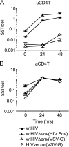
VSV-G-pseudotyped virions have impaired reverse transcription in unstimulated CD4+ T cells. Unstimulated CD4+ T cells (uCD4T) (A) or CD3/CD28 bead-stimulated CD4+ T cells (aCD4T) (B) (6, 43) were spinoculated with HIVvector(VSV-G), pNL4-3 (wtHIV), or pNL4-3Δenv pseudotyped with either VSV-G [HIVΔenv(VSV-G)] or HIV Env [HIVΔenv(HIV Env)] at a dose equivalent to ∼1 late reverse transcript per cell, as estimated in CEMss cells. The envelope of the HIV isolate LAI was used to pseudotype HIVΔenv particles. Late reverse transcription was measured by quantitative PCR using primers that detect the SST step of reverse transcription. The results were normalized to the peak level of reverse transcription in activated cells (∼1 SST/cell). Error bars represent the standard deviation of three measurements of reverse transcription. The figure is representative of two experiments. A slight increase in the level of reverse transcription by HIVΔenv(VSV-G) was detected by 48 h postinoculation. This slight increase in reverse transcription activity could be due to measurement variation at the lower limit of PCR detection (∼0.0001 to 0.005 SST/cell), minimal intravirion reverse transcription (29, 56, 65), or minimal and delayed reverse transcription. The apparent variation of background reverse transcription at 0 h postinoculation among viruses is likely due to variable contamination of the viral supernatants with plasmid DNA, which interferes with PCR detection of reverse transcription (see Supplement S2 in the supplemental material), and variable effectiveness of the treatment of the supernatants with DNase. Thus, the apparent minimal increase in reverse transcription from 0 h postinoculation by HIVΔenv(HIV Env) (filled triangles in panel B) is also likely due to contaminating plasmid DNA in the transfection supernatant.
FIG. 4.
FIG. 4.
VSV-G-pseudotyped virions do not fuse efficiently to unstimulated CD4+ T cells. Unstimulated CD4+ T cells (A and B) or CD3/CD28 bead-stimulated CD4+ T cells (C and D) were spinoculated with BlaM-Vpr-containing pNL4-3Δenv pseudotyped with VSV-G [HIVΔenv(VSV-G)] (A and C) or BlaM-Vpr-containing pNL4-3 (wtHIV) (B and D). The viral inocula were matched to have equivalent fusion activity levels (50% fusion) in CEMss cells. After spinoculation and induction of fusion at 37°C, the cells were loaded with the dye CCF2-AM, incubated at 25°C for 12 h, and analyzed by flow cytometry. Gates were set both by using the corresponding uninfected control and by setting the gate that measures viral fusion at ∼1% background. The figure is representative of two experiments.
FIG. 5.
FIG. 5.
Binding of VSV-G-pseudotyped virions to resting CD4+ T cells is less than that of wtHIV. Resting CD4+ T cells (CD25, CD69, and HLA DR negative) (rCD4T) or CD3/CD28 bead-stimulated CD4+ T cells (aCD4T) were spinoculated with HIVvector(VSV-G) or HIVvector(HIV Env) at a dose equivalent to ∼1 late reverse transcript per cell, as estimated in CEMss cells. The envelope of the HIV isolate LAI was used to pseudotype HIVvector particles. Viral binding was determined by measuring the level of cell-associated viral p24Gag immediately after inoculation. The level of cell-associated p24Gag was measured by ELISA. Error bars represent the standard deviation of a combined total of six (resting cells) or nine (activated cells) measurements of viral binding from three inoculations. Asterisks represent statistical significance at a P value of <0.01 as calculated by the Wilcoxon rank sum test. The figure is representative of three experiments.
FIG. 6.
FIG. 6.
Lentiviral vector particles pseudotyped with HIV envelope can transduce resting CD4+ T cells. Pure resting CD4+ T cells (rCD4T) (CD25, CD69, or HLA-DR negative) (A) or CD3/CD28 bead-stimulated CD4+ T cells (aCD4T) (B) were spinoculated with HIVvector(HIV Env) or HIVvector(VSV-G) at a dose equivalent to ∼1 late reverse transcript per cell, as estimated in stimulated CD4+ T cells. At the indicated time points postinoculation, integration was measured by Alu PCR. The graphs represent a combination of two experiments. Undetectable levels of integration were plotted directly on the x axis without error bars. Error bars represent the standard deviation of four to five combined measurements of integration. (C) Two different dilutions of HIVvector(HIV Env) were used to inoculate pure resting CD4+ T cells (rCD4T). The envelope of the HIV isolate LAI was used to pseudotype HIVvector particles. Integration was measured at 48 h postinfection by Alu PCR. The graph represents a combination of two (1:35 dilution) or three (1:7 dilution) experiments. Error bars represent the standard deviation of 5 (1:35 dilution) to 19 (1:7 dilution) combined measurements of integration.
FIG. 7.
FIG. 7.
Viral particles pseudotyped with HIV envelope do not induce expression of activation markers or cell cycle entry in resting CD4+ T cells. (A) Mock-infected or HIVvector(HIV Env)-infected pure resting CD4+ T cells (at ∼1 to 3 SST per cell in activated CD4+ T cells) were stained for CD4 and the cell activation markers CD25, CD69, and HLA-DR at various time points postinoculation. The gates for each time point were set using a fluorescence-minus-one control corresponding to each time point, similar to the method used for the experiment shown in Fig. 1A. (B) Mock-infected or HIVvector(HIV Env)-infected pure resting CD4+ T cells (at ∼1 to 3 SST per cell in activated CD4+ T cells) were stained with pyronin Y and 7-AAD to monitor cell cycle progression at 0, 4, and 24 h postinfection. The gates described in the legend of Fig. 1B were applied to establish the cell cycle stage.

References

    1. Adachi, A., H. E. Gendelman, S. Koenig, T. Folks, R. Willey, A. Rabson, and M. A. Martin. 1986. Production of acquired immunodeficiency syndrome-associated retrovirus in human and nonhuman cells transfected with an infectious molecular clone. J. Virol. 59284-291. - PMC - PubMed
    1. Agosto, L. M., J. J. Yu, J. Dai, R. Kaletsky, D. Monie, and U. O'Doherty. 2007. HIV-1 integrates into resting CD4+ T cells even at low inoculums as demonstrated with an improved assay for HIV-1 integration. Virology 36860-72. - PMC - PubMed
    1. Aiuti, A. 2002. Advances in gene therapy for ADA-deficient SCID. Curr. Opin. Mol. Ther. 4515-522. - PubMed
    1. Akkina, R. K., R. M. Walton, M. L. Chen, Q. X. Li, V. Planelles, and I. S. Chen. 1996. High-efficiency gene transfer into CD34+ cells with a human immunodeficiency virus type 1-based retroviral vector pseudotyped with vesicular stomatitis virus envelope glycoprotein G. J. Virol. 702581-2585. - PMC - PubMed
    1. Bukrinsky, M. I., T. L. Stanwick, M. P. Dempsey, and M. Stevenson. 1991. Quiescent T lymphocytes as an inducible virus reservoir in HIV-1 infection. Science 254423-427. - PMC - PubMed

Publication types

MeSH terms

Substances