. 2009 Aug;83(16):8021-31.
doi: 10.1128/JVI.00826-09. Epub 2009 Jun 3.

A complicated message: Identification of a novel PB1-related protein translated from influenza A virus segment 2 mRNA

Affiliations

A complicated message: Identification of a novel PB1-related protein translated from influenza A virus segment 2 mRNA

Helen M Wise et al. J Virol. 2009 Aug.

Abstract

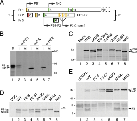
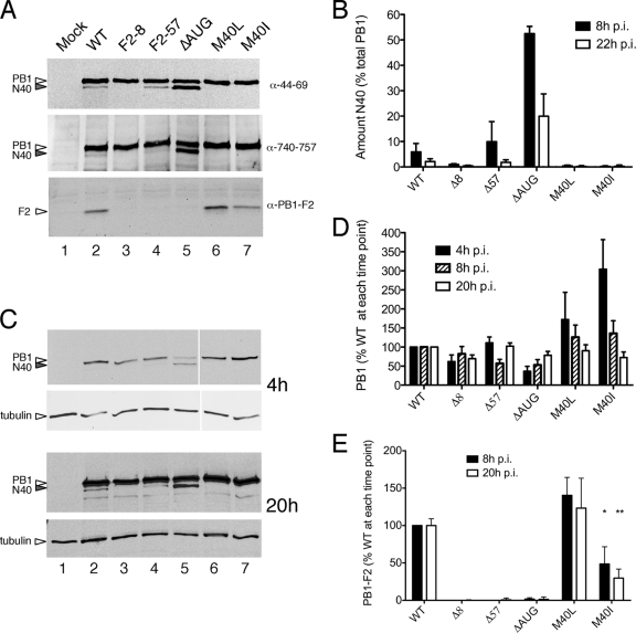
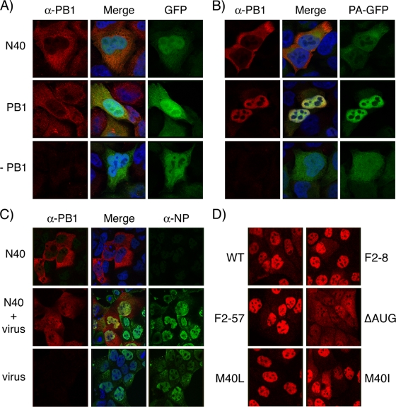
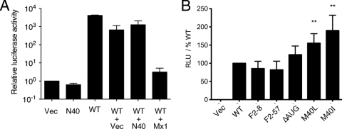
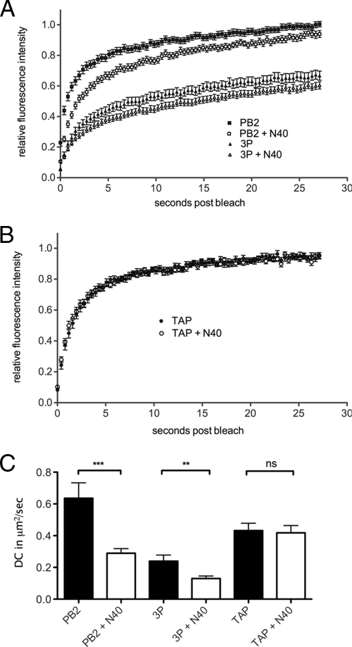
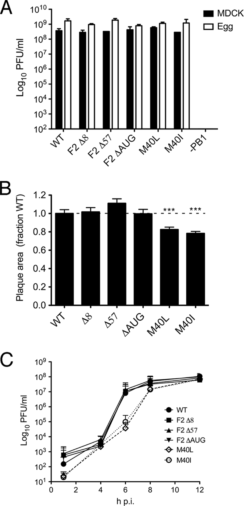
Influenza A virus segment 2 is known to encode two polypeptides in overlapping open reading frames: PB1, the polymerase, and PB1-F2, a proapoptotic virulence factor. We show that a third major polypeptide is synthesized from PB1 mRNA via differential AUG codon usage. PB1 codon 40 directs translation of an N-terminally truncated version of the polypeptide (N40) that lacks transcriptase function but nevertheless interacts with PB2 and the polymerase complex in the cellular environment. Importantly, the expression of N40, PB1-F2, and PB1 are interdependent, and certain mutations previously used to ablate PB1-F2 production affected N40 accumulation. Removal of the PB1-F2 AUG upregulated N40 synthesis, while truncating PB1-F2 after codon 8 (with a concomitant M40I change in PB1) abolished N40 expression. A virus lacking both N40 and PB1-F2 replicated normally. However, viruses that did not express N40 but retained an intact PB1-F2 gene overexpressed PB1 early in infection and replicated slowly in tissue culture. Thus, the influenza A virus proteome includes a 12th primary translation product that (similarly to PB1-F2) is nonessential for virus viability but whose loss, in particular genetic backgrounds, is detrimental to virus replication.


Figures

FIG. 1.
FIG. 1.
Expression of multiple protein species from IAV segment 2. (A) Diagram of the 5′ end of segment 2 mRNA. ORFs in the three reading frames are indicated by boxes. AUG codons are color coded according to the relative strength of their Kozak consensus (green = strong, −3A/G, +4G; amber = intermediate, −3A/G or + 4G; red = weak, −3U, +4U), and those that initiate identified polypeptides are indicated. (B) Detection of a minor PB1 species from infected cells. [35S]methionine-labeled lysate from MDCK cells infected (lanes I) or mock infected (lanes M) with Cambridge PR8 virus were immunoprecipitated with anti-PB1 V19, anti-PA V35, or anti-PB2 V28 as indicated and analyzed by SDS-PAGE and autoradiography in parallel with a radiolabeled in vitro translation reaction of WT segment 2 (in lane R, a lower exposure of the same track was used). (C) Lysates from cells infected (or mock infected) with the indicated panel of viruses, as well as in vitro-translated N40 were analyzed by Western blotting for PB1 with anti-PB1 V19 serum. (D) Aliquots of in vitro translation reactions programmed with the indicated plasmids (lane VOC [vector only control]) were analyzed by SDS-PAGE and autoradiography. (E) Lysates from 293T cells transfected with the indicated plasmids (pDUAL; empty vector control) were analyzed by SDS-PAGE and Western blotting with anti-PB1 MAb 10.4 (top panel) or anti-PB1-F2 C-terminal specific serum (bottom panel). The migration of molecular mass markers (in kilodaltons), as well as specified polypeptides, is indicated.
FIG. 2.
FIG. 2.
Expression of segment 2 polypeptides in virus-infected cells. MDCK cells were infected (or mock infected) with the panel of viruses as labeled, and cell lysates were obtained at various times p.i. (panel A, 8 h p.i.; panels C to E, as labeled) analyzed by Western blotting for the indicated polypeptides. Representative experiments are shown in panels A and C. (B, D, and E) The accumulation of the indicated polypeptides was quantified. The means ± the standard errors of the mean (SEM) of four independent experiments using two independently rescued virus stocks are plotted in panels B and D, while the data in panel E reflect the means and ranges of two independent experiments.
FIG. 3.
FIG. 3.
Intracellular localization of PB1-related polypeptides. (A and B) HeLa cells were transfected with plasmids encoding N40, PB1, GFP, or PA-GFP as indicated; fixed 24 h later; stained with anti-PB1 MAb 10.4; and imaged by confocal microscopy. (C) MDCK cells transfected with plasmids encoding N40 or (bottom row) empty vector were infected 24 h later with Dk/Sing virus and at 8 h p.i. were fixed and stained with anti-PB1 MAb 10.4 and anti-NP 2915 before imaging by confocal microscopy. (D) MDCK cells were infected with the indicated viruses, fixed, and stained with anti-PB1 MAb 10.4 at 8 h p.i. Confocal settings were kept consistent throughout each experiment.
FIG. 4.
FIG. 4.
Ability of segment 2 polypeptides to support IAV gene expression. 293T cells were transfected with plasmids encoding PB2, PA, NP, and a synthetic vRNA analogue containing a luciferase gene along with the indicated combinations of plasmids (WT [WT PB1]) and, 48 h later, the luciferase activity quantified. The data in panel A are the means ± the SEM of three independent experiments plotted relative to the activity seen in the absence of a PB1 gene, whereas the data in panel B are the means ± the SEM of a minimum of six independent experiments plotted as the percent activity seen with WT PB1 for each experiment. **, P < 0.01.
FIG. 5.
FIG. 5.
FRAP analysis of N40. 293T cells were transfected with plasmids encoding PB2-GFP or PB1-GFP, PA, and PB2 (3P) (A) or GFP-TAP (B) in combination with either N40 or an equivalent amount of empty vector, and the dynamics of the GFP-tagged proteins were examined by FRAP. Average (means ± the SEM) recovery curves from at least 32 individual cells are plotted in panel A and from at least 20 cells are plotted in panel B. (C) Mean plus SEM DC values are plotted. **, P < 0.01; ***, P < 0.001.
FIG. 6.
FIG. 6.
Growth properties of segment 2 mutant viruses. (A) Titers achieved at 48 h p.i. in MDCK cells infected at an MOI of 0.01 or at 48 h p.i. from eggs inoculated with 1,000 PFU are plotted. The means plus the ranges of two independent experiments are shown. (B) Mean plus SEM plaque areas in MDCK cells at 24 h p.i. are plotted. At least 150 plaques were measured from three separate experiments, and the data were normalized with respect to the mean plaque area of a matching WT virus. ***, P < 0.001. (C) Single cycle growth curves from MDCK cells infected at an MOI of 3. The means plus the SEM of four independent experiments using a minimum of two separate virus isolates are plotted.

Similar articles

Cited by

References

    1. Akkina, R. K., J. C. Richardson, M. C. Aguilera, and C. M. Yang. 1991. Heterogeneous forms of polymerase proteins exist in influenza A virus-infected cells. Virus Res. 1917-30. - PubMed
    1. Axelrod, D., D. E. Koppel, J. Schlessinger, E. Elson, and W. W. Webb. 1976. Mobility measurement by analysis of fluorescence photobleaching recovery kinetics. Biophys. J. 161055-1069. - PMC - PubMed
    1. Baigent, S. J., and J. W. McCauley. 2003. Influenza type A in humans, mammals and birds: determinants of virus virulence, host-range and interspecies transmission. Bioessays 25657-671. - PubMed
    1. Basler, C. F., and P. V. Aguilar. 2008. Progress in identifying virulence determinants of the 1918 H1N1 and the Southeast Asian H5N1 influenza A viruses. Antivir. Res. 79166-178. - PMC - PubMed
    1. Benfield, C. T., J. W. Lyall, G. Kochs, and L. S. Tiley. 2008. Asparagine 631 variants of the chicken Mx protein do not inhibit influenza virus replication in primary chicken embryo fibroblasts or in vitro surrogate assays. J. Virol. 827533-7539. - PMC - PubMed

Publication types

LinkOut - more resources