. 2009 Aug;83(16):8221-32.
doi: 10.1128/JVI.00576-09. Epub 2009 Jun 3.

Human papillomavirus type 16 infection of human keratinocytes requires clathrin and caveolin-1 and is brefeldin a sensitive

Affiliations

Human papillomavirus type 16 infection of human keratinocytes requires clathrin and caveolin-1 and is brefeldin a sensitive

Valerie Laniosz et al. J Virol. 2009 Aug.

Abstract

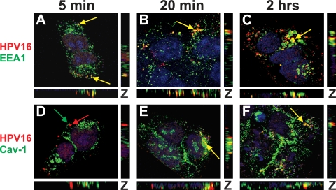
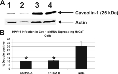
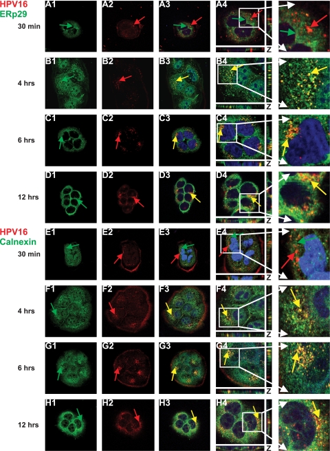
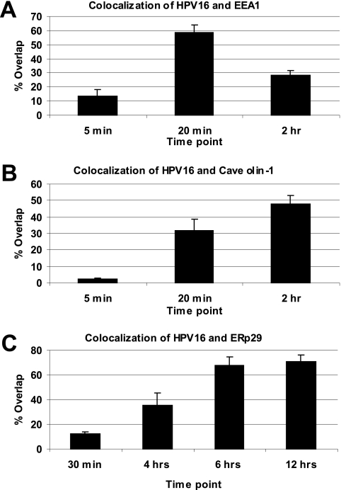
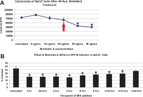
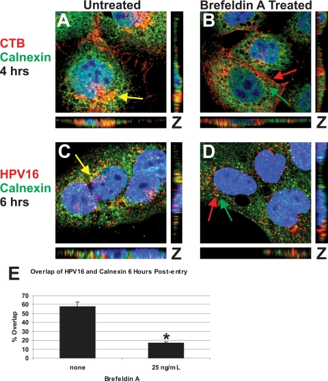
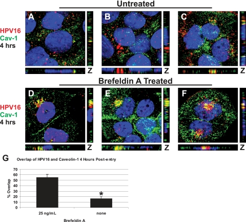
Human papillomavirus type 16 (HPV16) has been identified as being the most common etiological agent leading to cervical cancer. Despite having a clear understanding of the role of HPV16 in oncogenesis, details of how HPV16 traffics during infection are poorly understood. HPV16 has been determined to enter via clathrin-mediated endocytosis, but the subsequent steps of HPV16 infection remain unclear. There is emerging evidence that several viruses take advantage of cross talk between routes of endocytosis. Specifically, JCV and bovine papillomavirus type 1 have been shown to enter cells by clathrin-dependent endocytosis and then require caveolin-1-mediated trafficking for infection. In this paper, we show that HPV16 is dependent on caveolin-1 after clathrin-mediated endocytosis. We provide evidence for the first time that HPV16 infection is dependent on trafficking to the endoplasmic reticulum (ER). This novel trafficking may explain the requirement for the caveolar pathway in HPV16 infection because clathrin-mediated endocytosis typically does not lead to the ER. Our data indicate that the infectious route for HPV16 following clathrin-mediated entry is caveolin-1 and COPI dependent. An understanding of the steps involved in HPV16 sorting and trafficking opens up the possibility of developing novel approaches to interfere with HPV16 infection and reduce the burden of papillomavirus diseases including cervical cancer.


Figures

FIG. 1.
FIG. 1.
HPV16 traffics to early endosomes and overlaps with caveolin-1 by 20 min postentry. HaCaT cells infected with HPV16 reporter virions were stained with anti-L1 antibody H16.V5 (red) and either anti-EEA1 (A to C) (green) or anti-caveolin-1 (D to F) (green). HPV16 overlap with EEA1 was seen at 5 min (A), 20 min (B), and 2 h (C) (yellow arrows). HPV16 did not colocalize with caveolin-1 at 5 min (D) (red and green arrows). Colocalization of reporter virions and caveolin-1 was observed 20 min (E) and 2 h (F) postinfection (yellow arrows). The nucleus was stained using Topro-3 (blue). Images represent z stacks, showing the pattern of staining in the x, y, and z planes.
FIG. 2.
FIG. 2.
Caveolin-1 shRNA expression in HaCaT cells reduces HPV16 infection levels. (A) HaCaT cells were transfected with shRNA against caveolin-1 (Cav-1) in a plasmid containing a GFP reporter gene (lanes 1 and 2, shRNA A and shRNA B, respectively) or infected with a lentiviral vector containing GFP and siRNA against luciferase (lane 3). Lane 4 shows the lysates from HaCaT cells alone. Cell lysates were harvested for analysis by Western blotting after 48 h. The amount of caveolin-1 protein expression was determined by blotting with an anti-caveolin-1 antibody. Actin was used as a loading control. (B) Infection in HaCaT cells expressing shRNA A, shRNA B, or siRL was assessed after 48 h. Cells were harvested, and the percentage of cells that were GFP positive (expressing shRNA or siRL) and also DsRed positive (infected) was measured by flow cytometry as the percentage of double-positive cells. The drop in infection in caveolin-1 knockdown cells was determined to be significant (*, P < 0.002). Each bar represents the average of data from three trials for which the standard deviations are shown by error bars.
FIG. 3.
FIG. 3.
Colocalization of HPV16 and ER markers by 4 h after internalization. HaCaT cells infected with HPV16 reporter virions (red) were stained for ERp29 (A to D) (green) or calnexin (E to H) (green). (A) Overlap between HPV16 and ERp29 was not seen at 30 min (A3 and 4) (red and green arrows). (B to D) Colocalization of HPV16 and ERp29 was observed at 4 h (B3 and 4), 6 h (C3 and 4), and 12 h (D3 and 4) (yellow arrows). (E) HPV16 and calnexin did not overlap at 30 min postentry (E3 and 4, red and green arrows). (F to H) At 4 h (F3 and 4), 6 h (G3 and 4), and 12 h (H3 and 4) after HPV16 internalization, virions colocalized with calnexin (yellow arrows). Topro-3 was used to identify the nucleus (blue). z-stacked images show nine 0.5-μm slices in the x, y, and z planes. Enlarged sections of the z-stacked images are shown to the right.
FIG. 4.
FIG. 4.
Quantification of overlap between HPV16 reporter virions and intracellular markers. Immunofluorescence images were analyzed to quantify the colocalization of HPV16 and intracellular markers. (A) Percentage of HPV16 virions that colocalized with the early endosome marker EEA1 at 5 min, 20 min, and 2 h. (B) Percentage of HPV16 virions that colocalized with caveolin-1, a marker for vesicles of the caveolar pathway, at 5 min, 20 min, and 2 h. (C) The colocalization of HPV16 reporter virions and the ER marker ERp29 was quantified. The percent overlap at 30 min, 4 h, 6 h, and 12 h shows a time-dependent increase in overlap between reporter virions and the ER. Analysis was performed with a minimum of three separate images per time point. Error bars represent the standard deviations from the means.
FIG. 5.
FIG. 5.
BFA treatment inhibits HPV16 infection in a time-dependent manner. (A) BFA was added to HaCaT cells at increasing concentrations. Forty-eight hours later, cell viability was assessed as a function of the level of ATP. Doses of BFA of 35 and 50 ng/ml were found to be toxic to HaCaT cells (*, P < 0.005). A total of 25 ng/ml of BFA (red arrow) was used in time course and immunofluorescence studies. Error bars show the standard deviations from the means of data from a minimum of three experiments. (B) BFA was added to HaCaT cells at various time points in relation to HPV16 internalization. HPV16 infection was reduced to a statistically significant level in cells treated with BFA until the addition of BFA 24 h after HPV16 entry (*, P < 0.04). Error bars show the standard deviations of data from three experiments in which 10,000 cells were analyzed by flow cytometry for reporter gene expression to obtain the percentage of infected cells.
FIG. 6.
FIG. 6.
BFA blocks cholera toxin B and HPV16 trafficking to the ER. (A and B) Cholera toxin B (CTB) (red) overlap with the ER marker calnexin (green) at 4 h was assessed. (A) Colocalization was seen between cholera toxin B and calnexin in untreated HaCaT cells (yellow arrow). (B) The amount of cholera toxin B trafficking to the ER was greatly reduced in the presence of BFA (red and green arrows). (C and D) HPV16 was internalized for 6 h, and cells were stained with anti-L1 antibody H16.V5 (red) and anti-calnexin (green). (C) Untreated HaCaT cells showed colocalization between HPV16 and calnexin and prominent perinuclear accumulation (yellow arrow). (D) BFA treatment resulted in a loss of this overlap (red and green arrows). Topro-3 (blue) was used for the detection of the nucleus. z stack pictures are shown, depicting the image in the x, y, and z planes. (E) The percentage of HPV16 reporter virions overlapping with calnexin in untreated (none) or BFA-treated (25 ng/ml) HaCaT cells was quantified. The reduction in the overlap between HPV16 and calnexin in BFA-treated cells was statistically significant compared to the overlap in untreated cells (*, P < 0.001). At least three separate images were quantified, and the error bars represent the standard deviations from means.
FIG. 7.
FIG. 7.
Postcaveolar vesicle trafficking of HPV16 is BFA sensitive. HPV16 reporter virions internalized into HaCaT cells for 4 h in the absence (A to C) or in the presence (D to F) of BFA were detected with H16.V5 antibody (red). Anti-caveolin-1 antibody was used to identify caveolar vesicles (green). (A to C) In untreated cells, HPV16 overlap with caveolin-1 was minimal. (D to F) Compared to untreated cells, BFA treatment of HaCaT cells led to increased colocalization between HPV16 and caveolin-1 (yellow). Images represent z stacks showing the cells in all three planes. (G) Overlap of HPV16 reporter virions and caveolin-1 in BFA-treated (25 ng/ml) and untreated (none) HaCaT cells was quantified. The increased colocalization of HPV16 and caveolin-1 in BFA-treated cells compared to untreated cells was statistically significant (*, P < 0.002). Error bars represent the standard deviations from the means after analysis of at least three separate images.

Similar articles

Cited by

References

    1. Abban, C. Y., N. A. Bradbury, and P. I. Meneses. 2008. HPV16 and BPV1 infection can be blocked by the dynamin inhibitor dynasore. Am. J. Ther. 15304-311. - PMC - PubMed
    1. Ahluwalia, N., J. J. Bergeron, I. Wada, E. Degen, and D. B. Williams. 1992. The p88 molecular chaperone is identical to the endoplasmic reticulum membrane protein, calnexin. J. Biol. Chem. 26710914-10918. - PubMed
    1. Anderson, H. A., Y. Chen, and L. C. Norkin. 1996. Bound simian virus 40 translocates to caveolin-enriched membrane domains, and its entry is inhibited by drugs that selectively disrupt caveolae. Mol. Biol. Cell 71825-1834. - PMC - PubMed
    1. Bergeron, J. J., M. B. Brenner, D. Y. Thomas, and D. B. Williams. 1994. Calnexin: a membrane-bound chaperone of the endoplasmic reticulum. Trends Biochem. Sci. 19124-128. - PubMed
    1. Bethune, J., F. Wieland, and J. Moelleken. 2006. COPI-mediated transport. J. Membr. Biol. 21165-79. - PubMed

Publication types

MeSH terms

LinkOut - more resources