

Prions are secreted in milk from clinically normal scrapie-exposed sheep
- PMID: 19494004
- PMCID: PMC2715765
- DOI: 10.1128/JVI.00051-09
Prions are secreted in milk from clinically normal scrapie-exposed sheep
Abstract
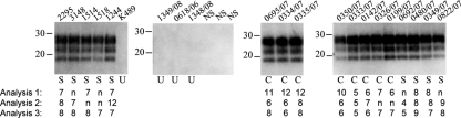
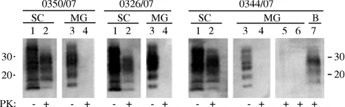
The potential spread of prion infectivity in secreta is a crucial concern for prion disease transmission. Here, serial protein misfolding cyclic amplification (sPMCA) allowed the detection of prions in milk from clinically affected animals as well as scrapie-exposed sheep at least 20 months before clinical onset of disease, irrespective of the immunohistochemical detection of protease-resistant PrP(Sc) within lymphoreticular and central nervous system tissues. These data indicate the secretion of prions within milk during the early stages of disease progression and a role for milk in prion transmission. Furthermore, the application of sPMCA to milk samples offers a noninvasive methodology to detect scrapie during preclinical/subclinical disease.
Figures
Similar articles
-
The oral secretion of infectious scrapie prions occurs in preclinical sheep with a range of PRNP genotypes.J Virol. 2012 Jan;86(1):566-71. doi: 10.1128/JVI.05579-11. Epub 2011 Oct 19. J Virol. 2012. PMID: 22013047 Free PMC article.
-
PrP(Sc) detection and infectivity in semen from scrapie-infected sheep.J Gen Virol. 2012 Jun;93(Pt 6):1375-1383. doi: 10.1099/vir.0.038802-0. Epub 2012 Feb 8. J Gen Virol. 2012. PMID: 22323531
-
Evidence of scrapie transmission to sheep via goat milk.BMC Vet Res. 2016 Sep 17;12:208. doi: 10.1186/s12917-016-0807-4. BMC Vet Res. 2016. PMID: 27640200 Free PMC article.
-
Transition of the prion protein from a structured cellular form (PrPC ) to the infectious scrapie agent (PrPSc ).Protein Sci. 2019 Dec;28(12):2055-2063. doi: 10.1002/pro.3735. Epub 2019 Oct 25. Protein Sci. 2019. PMID: 31583788 Free PMC article. Review.
-
Molecular biology of prions causing infectious and genetic encephalopathies of humans as well as scrapie of sheep and BSE of cattle.Dev Biol Stand. 1991;75:55-74. Dev Biol Stand. 1991. PMID: 1686599 Review.
Cited by
-
The oral secretion of infectious scrapie prions occurs in preclinical sheep with a range of PRNP genotypes.J Virol. 2012 Jan;86(1):566-71. doi: 10.1128/JVI.05579-11. Epub 2011 Oct 19. J Virol. 2012. PMID: 22013047 Free PMC article.
-
Accelerated shedding of prions following damage to the olfactory epithelium.J Virol. 2012 Feb;86(3):1777-88. doi: 10.1128/JVI.06626-11. Epub 2011 Nov 30. J Virol. 2012. PMID: 22130543 Free PMC article.
-
New generation QuIC assays for prion seeding activity.Prion. 2012 Apr-Jun;6(2):147-52. doi: 10.4161/pri.19430. Epub 2012 Apr 1. Prion. 2012. PMID: 22421206 Free PMC article. Review.
-
Detection of prions in oocytes and ovaries of ewes naturally infected with classical scrapie.Vet Res. 2025 Apr 10;56(1):79. doi: 10.1186/s13567-025-01512-0. Vet Res. 2025. PMID: 40211373 Free PMC article.
-
Extraneural manifestations of prion infection in GPI-anchorless transgenic mice.Virology. 2011 Mar 1;411(1):1-8. doi: 10.1016/j.virol.2010.12.012. Epub 2011 Jan 11. Virology. 2011. PMID: 21227476 Free PMC article.
References
-
- Castilla, J., P. Saá, and C. Soto. 2005. Detection of prions in blood. Nat. Med. 11982-985. - PubMed
-
- Didier, A., R. Gebert, R. Dietrich, M. Schweiger, M. Gareis, E. Märtlbauer, and W. M. Amselbruber. 2008. Cellular prion protein in mammary gland and milk fractions of domestic ruminants. Biochem. Biophys. Res. Commun. 369841-844. - PubMed
-
- Fox, K. A., J. E. Jewell, E. S. Williams, and M. W. Miller. 2006. Patterns of PrPCWD accumulation during the course of chronic wasting disease infection in orally inoculated mule deer (Odicoileus hemionus). J. Gen. Virol. 873451-3461. - PubMed
MeSH terms
Substances
LinkOut - more resources
Full Text Sources
Research Materials
