. 2009 Aug;83(16):8141-52.
doi: 10.1128/JVI.02116-08. Epub 2009 Jun 3.

Activation of intracellular signaling pathways by the murine cytomegalovirus G protein-coupled receptor M33 occurs via PLC-{beta}/PKC-dependent and -independent mechanisms

Affiliations

Activation of intracellular signaling pathways by the murine cytomegalovirus G protein-coupled receptor M33 occurs via PLC-{beta}/PKC-dependent and -independent mechanisms

Joseph D Sherrill et al. J Virol. 2009 Aug.

Abstract

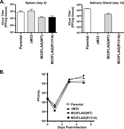
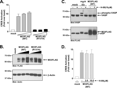
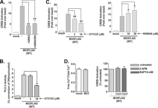
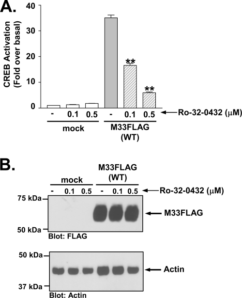
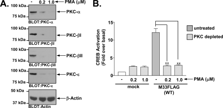
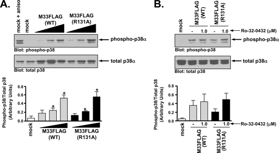
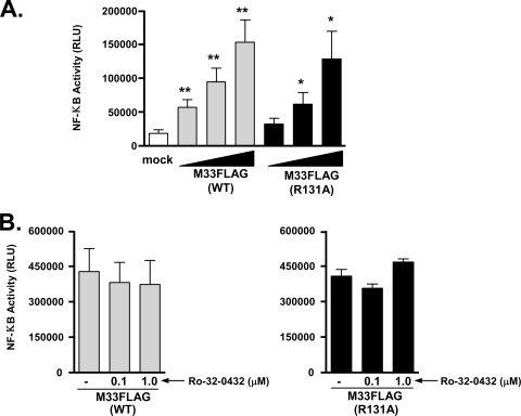
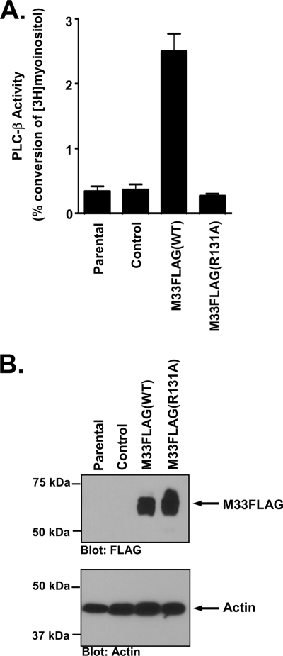
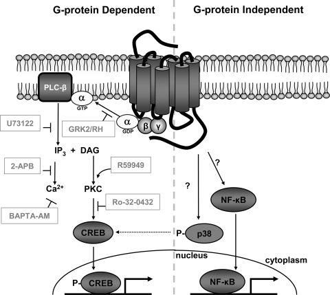
The presence of numerous G protein-coupled receptor (GPCR) homologs within the herpesvirus genomes suggests an essential role for these genes in viral replication in the infected host. Such is the case for murine cytomegalovirus (MCMV), where deletion of the M33 GPCR or replacement of M33 with a signaling defective mutant has been shown to severely attenuate replication in vivo. In the present study we utilized a genetically altered version of M33 (termed R131A) in combination with pharmacological inhibitors to further characterize the mechanisms by which M33 activates downstream signaling pathways. This R131A mutant of M33 fails to support salivary gland replication in vivo and, as such, is an important tool that can be used to examine the signaling activities of M33. We show that M33 stimulates the transcription factor CREB via heterotrimeric G(q/11) proteins and not through promiscuous coupling of M33 to the G(s) pathway. Using inhibitors of signaling molecules downstream of G(q/11), we demonstrate that M33 stimulates CREB transcriptional activity in a phospholipase C-beta and protein kinase C (PKC)-dependent manner. Finally, utilizing wild-type and R131A versions of M33, we show that M33-mediated activation of other signaling nodes, including the mitogen-activated protein kinase family member p38alpha and transcription factor NF-kappaB, occurs in the absence of G(q/11) and PKC signaling. The results from the present study indicate that M33 utilizes multiple mechanisms to modulate intracellular signaling cascades and suggest that signaling through PLC-beta and PKC plays a central role in MCMV pathogenesis in vivo.


Figures

FIG. 1.
FIG. 1.
M33FLAG(R131A) recombinant virus fails to persist in the salivary glands. (A) Female BALB/c Mice were infected intraperitoneally with 5 × 10⁵ PFU of K181 BAC-derived parental, ΔM33, M33FLAG(WT), or M33FLAG(R131A) viruses. Groups of three mice were sacrificed on day 4 or day 14 postinfection, and their spleens and salivary glands were harvested. Tissues were homogenized, and virus titers were assessed by plaque assay on NIH 3T3 cells. Virus titers from spleens isolated at 4 days postinfection (left panel) or from salivary glands isolated at 14 days postinfection (right panel) are depicted graphically. (B) The ability of each of the BAC-derived viruses to replicate in salivary epithelial cells was also assessed in the salivary gland cell line SGC-1. Multistep growth curves were performed by infecting subconfluent SGC-1 cells at a multiplicity of infection of 0.1. Samples of tissue culture supernatant were taken at days 1, 3, and 5 postinfection, and virus titers were determined by plaque assay. The data represent the means of three independent experiments.
FIG. 2.
FIG. 2.
M33 activates CREB independent of Gs coupling. (A) HEK293 cells were transiently transfected with empty vector (mock) or 50, 100, or 250 ng of M33FLAG(WT) or M33FLAG(R131A), and CREB-driven luciferase transcription was measured and normalized to Renilla luciferase. The data represent the means ± the standard errors (SE) of four independent experiments performed in duplicate. (B) The expression of M33FLAG(WT) and M33-FLAG(R131A) in similarly transfected HEK293 cells was assessed by Western blotting. (C) PKA activity, as assessed by the phosphorylation status of the PKA substrate VASP, was assessed by Western blotting in HEK293 cells stably transfected with empty vector (mock) or M33 (M33FLAG). Cells were also transfected with FLAG-VASP. Control cells (mock) unstimulated or stimulated with the β-adrenergic agonist isoproterenol (1 μM for 5 min) with or without pretreatment with the PKA inhibitor H-89 were included to validate the sensitivity and specificity of the assay. The phosphorylation status of VASP was assessed by Western blotting with an anti-VASP polyclonal antibody (upper panel). Expression of M33FLAG was confirmed by Western blotting with an anti-FLAG polyclonal antibody (lower panel). Note that the bands in the non-M33 transfected cells represent phosphorylated or nonphosphorylated VASP which, like M33, is also FLAG tagged. The immunoblots shown are representative of at least four independent experiments. (D) HEK293 cells were transiently transfected with empty vector (mock) or M33FLAG(WT) (250 ng of each), and CREB-driven luciferase transcription normalized to Renilla luciferase was measured in the presence or absence of the PKA inhibitor H-89 (2.0 or 10 μM for 24 h). The data represent the means ± the SE of three independent experiments performed in duplicate.
FIG. 3.
FIG. 3.
M33-induced CREB activation occurs through the Gq/11/PLC-β pathway. (A) HEK293 cells were transiently transfected with empty vector (mock) or M33FLAG(WT) (250 ng) with or without GRK2 (250 ng) or the Gαq/11-binding domain of GRK2 (GRK2/RH) (250 ng), and CREB-driven luciferase transcription was measured and normalized to Renilla luciferase. The data represent the means ± the SE of three independent experiments performed in duplicate. (B) HEK293 cells were transiently transfected with empty vector (mock) or M33FLAG(WT) (1.25 μg), and accumulated inositol phosphates were measured. M33-expressing cells were left untreated or treated with the PLC-β inhibitor U73122 (3 and 10 μM) for 24 h prior to assay. The data represent the means ± the SE of three independent experiments performed in duplicate. (C) CREB-driven luciferase activity normalized to Renilla luciferase was measured in HEK293 cells transiently transfected with M33FLAG(WT) (250 ng) untreated or treated with the PLC-β inhibitor U73122 (3 and 10 μM for 24 h, left panel) or the diacyglycerol kinase inhibitor R59949 (10 and 30 μM for 24 h, right panel). The data represent the means ± the SE of five independent experiments performed in duplicate. (D) Cells transiently transfected with empty vector or M33 (1.25 μg) were assessed for intracellular Ca2+ levels using Fluo-4 NW (left panel). CREB-driven luciferase activity normalized to Renilla luciferase was measured in HEK293 cells transiently transfected with M33FLAG(WT) (250 ng) untreated or treated with the IP3 channel inhibitor 2-APB (20 μM for 24 h) or the calcium chelator BAPTA-AM (30 μM for 24 h, right panel). The data represent the means ± the SE of three independent experiments performed in duplicate. *, P ≤ 0.05; **, P ≤ 0.005.
FIG. 4.
FIG. 4.
M33-induced CREB activation is mediated by PKC. (A) HEK293 cells stably transfected with empty vector (mock) or M33FLAG were treated with the PKC inhibitor Ro-32-0432 (0.1 or 0.5 μM for 24 h) and CREB-driven luciferase activity was measured and normalized to Renilla luciferase. (B) Expression of M33FLAG was confirmed by Western blotting with an anti-FLAG polyclonal antibody. The data represent the means ± the SE of three independent experiments performed in duplicate. **, P ≤ 0.005.
FIG. 5.
FIG. 5.
M33-induced CREB activation is defective in PKC-depleted cells. (A) HEK293 cells were treated overnight with the phorbol ester PMA to deplete endogenous PKCs. HEK293 cells express several isoforms of PKC including PKC-α, PKC-β1, PKC-β2, and PKC-ɛ and Western blotting of cells treated with 0.2 or 1.0 μM PMA confirmed that these conditions resulted in >90% depletion of endogenous PKCs. (B) After depletion of endogenous PKCs, untreated or treated cells were transiently transfected with empty vector (mock) or M33FLAG(WT) (250 ng) and CREB-driven luciferase activity was measured and normalized to Renilla luciferase. The data represent the mean ± the SE of four independent experiments performed in duplicate. **, P ≤ 0.005.
FIG. 6.
FIG. 6.
M33 activates the MAPK p38 independent of Gq/11/PKC signaling. (A) HEK293 cells were cotransfected with M33FLAG(WT) or M33FLAG(R131A) (0.25, 0.50, and 1.25 μg) and FLAG-p38α (0.50 μg) and p38 phosphorylation was assessed by Western blotting (upper panel). As a positive control, mock-transfected cells were treated with the stress response activator anisomycin. The results are presented graphically and represent the means ± the SE of four independent experiments (lower panel). (B) Phosphorylated p38 levels in HEK293 cells expressing M33FLAG(WT) or M33FLAG(R131A) (1.25 μg of each) and FLAG-p38α (0.5 μg) were assessed in the presence or absence of the PKC inhibitor Ro-32-0432 (upper panel). The results are presented graphically and represent the means ± the SE of three independent experiments (lower panel). *, P ≤ 0.05.
FIG. 7.
FIG. 7.
Stimulation of NF-κB transcriptional activity by M33 is independent of Gq/11/PKC signaling. (A) HEK293 cells were transfected with empty pCDNA3 (mock) or increasing amounts of M33(WT) or M33(R131A) (10, 50, and 250 ng), and NF-κB-driven luciferase activity was measured and normalized to Renilla luciferase. The data represent the means ± the SE of five independent experiments performed in duplicate. (B) HEK293 cells transfected with M33(WT) or M33(R131A) (250 ng of each) were pretreated with the PKC inhibitor Ro-32-0432 (1.0 μM for 24 h) and NF-κB-driven luciferase activity was measured and normalized to Renilla luciferase. The data represent the means ± the SE of three independent experiments performed in duplicate. RLU, relative light units. *, P ≤ 0.05; **, P ≤ 0.005.
FIG. 8.
FIG. 8.
M33 activates the Gq/11/PLC-β pathway in salivary epithelial cells. (A) SGC1 salivary epithelial cells were transduced with the retroviruses MIGR1, MIGR1-M33FLAG(WT), or MIGR1-M33FLAG(R131A). PLC-β activity was then measured in each cell line and is expressed as the percent conversion of input myoinositol into inositol phosphates. The data represent the means ± the SE of five independent experiments performed in duplicate. (B) Expression of M33FLAG and M33FLAG(R131A) in the transduced SGC1 cells was confirmed by Western blotting with an anti-FLAG polyclonal antibody (upper panel).
FIG. 9.
FIG. 9.
G protein-dependent and G protein-independent signaling activities of M33. Use of pharmacological and genetic inhibitors at multiple steps along the Gq/11 signaling pathway reveals that CREB activation by the MCMV GPCR M33 is mediated by coupling to heterotrimeric Gq/11 proteins and PLC-β activity. Moreover, while not affected by IP3 and Ca2+ levels, CREB activity is dependent on PKC activity and is augmented by increased levels of DAG. Conversely, M33 stimulation of p38 (which may partially contribute to M33-induced CREB activity) and NF-κB occurs in the absence of Gq/11 signaling through an as-yet-defined mechanism(s).

Similar articles

Cited by

References

    1. Ahn, S., S. K. Shenoy, H. Wei, and R. J. Lefkowitz. 2004. Differential kinetic and spatial patterns of beta-arrestin and G protein-mediated ERK activation by the angiotensin II receptor. J. Biol. Chem. 27935518-35525. - PubMed
    1. Alewijnse, A. E., H. Timmerman, E. H. Jacobs, M. J. Smit, E. Roovers, S. Cotecchia, and R. Leurs. 2000. The effect of mutations in the DRY motif on the constitutive activity and structural instability of the histamine H(2) receptor. Mol. Pharmacol. 57890-898. - PubMed
    1. Azzi, M., P. G. Charest, S. Angers, G. Rousseau, T. Kohout, M. Bouvier, and G. Pineyro. 2003. Beta-arrestin-mediated activation of MAPK by inverse agonists reveals distinct active conformations for G protein-coupled receptors. Proc. Natl. Acad. Sci. USA 10011406-11411. - PMC - PubMed
    1. Bais, C., B. Santomasso, O. Coso, L. Arvanitakis, E. G. Raaka, J. S. Gutkind, A. S. Asch, E. Cesarman, M. C. Gershengorn, and E. A. Mesri. 1998. G-protein-coupled receptor of Kaposi's sarcoma-associated herpesvirus is a viral oncogene and angiogenesis activator. Nature 39186-89. - PubMed
    1. Beisser, P. S., G. Grauls, C. A. Bruggeman, and C. Vink. 1999. Deletion of the R78 G protein-coupled receptor gene from rat cytomegalovirus results in an attenuated, syncytium-inducing mutant strain. J. Virol. 737218-7230. - PMC - PubMed

Publication types

MeSH terms

LinkOut - more resources