. 2009 Aug;83(16):7842-9.
doi: 10.1128/JVI.00309-09. Epub 2009 Jun 3.

The nucleoprotein of lymphocytic choriomeningitis virus facilitates spread of persistent infection through stabilization of the keratin network

Affiliations

The nucleoprotein of lymphocytic choriomeningitis virus facilitates spread of persistent infection through stabilization of the keratin network

Martina Labudova et al. J Virol. 2009 Aug.

Abstract

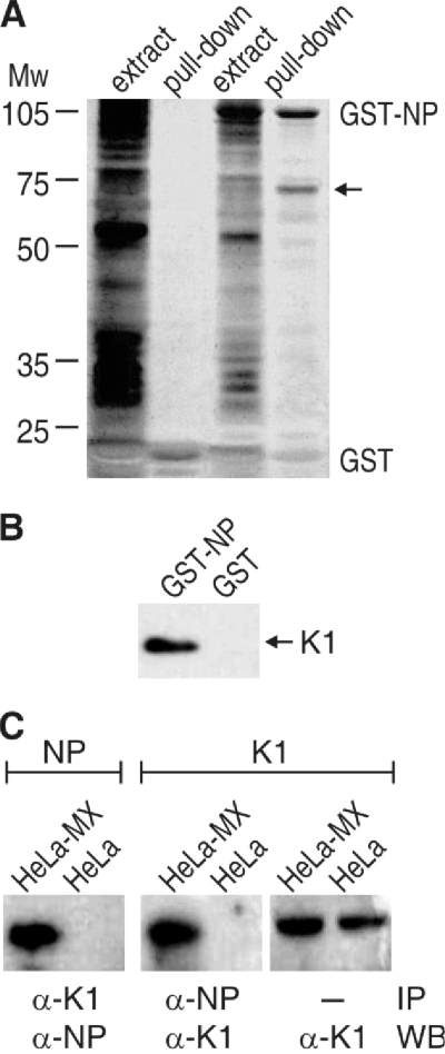
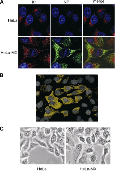
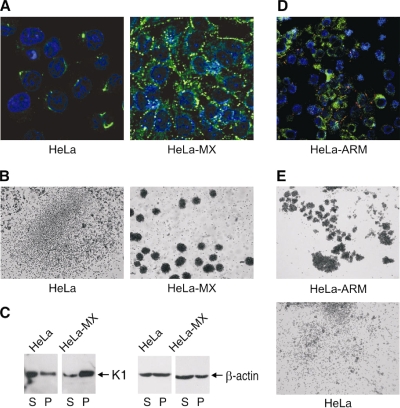
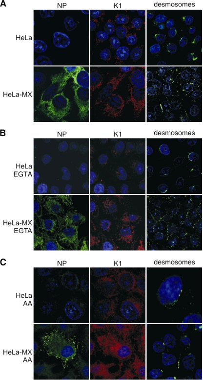
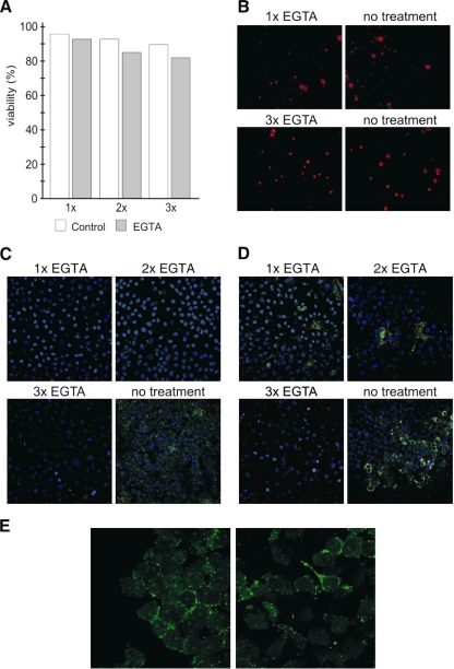
Lymphocytic choriomeningitis virus (LCMV) is a prototypic arenavirus containing a bisegmented single-stranded RNA genome with an ambisense coding strategy. MX is a noncytolytic LCMV strain with an in vitro host range restricted to only few cell lines. MX LCMV spreads via cell-cell contacts and causes persistent infection with high production of viral nucleoprotein (NP). Using a proteomic approach, we identified keratin 1 (K1), an intermediate filament network component, as a binding partner of the viral NP. The functional significance of this interaction has been examined by chemical disruption of the keratin network, resulting in a reduced spread of MX LCMV in HeLa cells. However, K1 disassembly was considerably lower in MX LCMV-infected cells than in noninfected counterparts, indicating that NP can stabilize the keratin network and thereby support the integrity of cytoskeleton. The presence of NP also resulted in increased formation of desmosomes and stronger cell-cell adhesion. Similar effects were observed in HeLa cells persistently infected with LCMV strain Armstrong. Our findings suggest that the keratin network is important for the intercellular transmission of persistent LCMV infection in epithelial cells and show that the virus can actively facilitate its own intercellular spread through the interaction between the viral NP and K1 and stimulation of cell-cell contacts.


Figures

FIG. 1.
FIG. 1.
Identification of K1 as an interacting partner of MX LCMV NP. (A) Pull-down assay. GST-NP fusion protein and GST were immobilized on glutathione-S Sepharose and incubated with extracts from HeLa cells. Proteins bound to GST-NP and GST were separated by SDS-PAGE, and whole extracts were loaded for the control of input. A prominent band corresponding to 70 kDa was detected among GST-NP pulled-down proteins (arrow) and identified by sequencing as K1. (B) Pull-down assay combined with Western blotting and immunodetection of K1. (C) Immunoprecipitation combined with Western blotting was performed as a proof of interaction between virus NP and K1. The antibodies used for each step are indicated at the bottom.
FIG. 2.
FIG. 2.
The K1 network in infected versus noninfected HeLa cells. (A) Immunofluorescence detection of K1 in HeLa-MX cells in comparison with noninfected HeLa controls and colocalization with MX LCMV NP using K1-specific (red) and NP-specific (green) MAbs. Cell nuclei were stained with DAPI (blue). HeLa-MX cells containing NP display a higher content and better organization of K1. Original magnification, ×63. (B) Three-dimensional colocalization map of NP and K1 signals obtained from double-staining confocal analysis of mixed HeLa and HeLa-MX monolayer. The figure shows only the sites where NP and K1 overlap in MX LCMV-infected cells (Pearson's coefficient of colocalization corresponds to 0.704). The colocalization signal is absent from noninfected cells containing only K1 and no NP. (C) Phase-contrast micrographs show a more prominent keratin network in MX LCMV-infected cells.
FIG. 3.
FIG. 3.
Formation of desmosomes in MX LCMV-infected HeLa cells. (A) Immunofluorescence analysis of desmosomes using the specific MAb ZK-31 (green) revealed a lack of desmosomal signal in noninfected HeLa cells. In contrast, HeLa-MX cells showed strong staining signals with a pattern typical for desmosomes. Cell nuclei were stained with DAPI (blue). (B) HeLa-MX cells also exhibited increased cell adhesion capacity in an assay based on spontaneous aggregation of cells first grown in a monolayer and then brought to single-cell suspension, placed on a nonadhesive surface, and rotated overnight on a gyratory shaker. (C) K1 assembly in soluble (S) and pellet (P) fractions of the cell extracts was analyzed by Western blotting. β-Actin was detected as a loading control. (D) Double-staining immunofluorescence analysis of viral NP (green) and desmosomes (red) in HeLa cells persistently infected with LCMV strain Armstrong. Infected cells show a clear desmosomal signal which is absent from noninfected counterparts. (E) An aggregation assay of infected HeLa-ARM cells compared to noninfected control HeLa cells confirmed increased clustering of infected cells.
FIG. 4.
FIG. 4.
Effect of keratin network disassembly on distribution of NP, K1, and desmosomes, showing immunofluorescence analysis of control HeLa and HeLa-MX cells (A) and of cells treated with the inhibitors of intermediate filaments EGTA (B) and acrylamide (AA) (C). The cells were treated for 15 min with 2 mM EGTA and for 8 h with 5 mM AA, and then they were fixed and stained with antibodies specific for NP (green), K1 (red), and desmosomes (green). Cell nuclei were stained with DAPI (blue). Both inhibitors caused a loss or reduction of K1 and desmosomal staining signals in the noninfected cells, whereas these signals were relatively well preserved in the infected cells. Original magnification, ×63.
FIG. 5.
FIG. 5.
Effect of keratin network disassembly and loss of cell-cell contacts on MX LCMV spread in HeLa cells. Immunofluorescence analysis of HeLa cells mixed with HeLa-MX cells in a 10:1 ratio was used to detect MX LCMV spread. (A and B) The mixed cell monolayers were treated with EGTA, and the viability of cells was assessed by flow cytometric analysis (A) and by fluorescence microscopy of cells stained with propidium iodide (B) as described in Materials and Methods. (C and D) For analysis of MX LCMV spread, the cells were stained with desmosome (C)- and NP (D)-specific antibodies. EGTA was added to cells and left for 15 min once, twice, or three times, with 24-h recovery periods between the treatments. Untreated cells were maintained in parallel. In the absence of EGTA treatment, clusters of neighboring cells show the presence of desmosomes and NP, suggesting that the virus was spread via cell-cell contacts. In contrast, virtually no desmosomes are visible in EGTA-treated cells, and the NP signal is either confined to few isolated cells or completely absent from the monolayer. Original magnification, ×10. (E) In order to show that LCMV spread is affected by disruption of intercellular contacts (as a consequence of disruption of the keratin network), we stained nonfixed cells with NP-specific antibody. Because antibody cannot cross the membranes of living cells, it could bind only to NP present on the cell surface as a component of minimal infection units. The figure shows that NP can be detected on the cell surface and that the signal is concentrated in the areas of intercellular contacts. On the other hand, it is not visible when cell-cell contacts are missing.

References

    1. Amman, B. R., B. I. Pavlin, C. G. Albariño, J. A. Comer, B. R. Erickson, J. B. Oliver, T. K. Sealy, M. J. Vincent, S. T. Nichol, C. D. Paddock, A. J. Tumpey, K. D. Wagoner, R. D. Glauer, K. A. Smith, K. A. Winpisinger, M. S. Parsely, P. Wyrick, C. H. Hannafin, U. Bandy, S. Zaki, P. E. Rollin, and T. G. Ksiazek. 2007. Pet rodents and fatal lymphocytic choriomeningitis in transplant patients. Emerg. Infect. Dis. 13719-725. - PMC - PubMed
    1. Buchmeier, M. J. 2002. Arenaviruses: protein structure and function. Curr. Top. Microbiol. Immunol. 262159-173. - PubMed
    1. Chidgey, M., and C. Dawson. 2007. Desmosomes: a role in cancer? Br. J. Cancer 961783-1787. - PMC - PubMed
    1. Diefenbach, R. J., M. Miranda-Saksena, E. Diefenbach, D. J. Holland, R. A. Boadle, P. J. Armati, and A. L. Cunningham. 2002. Herpes simplex virus tegument protein US11 interacts with conventional kinesin heavy chain. J. Virol. 763282-3291. - PMC - PubMed
    1. Dylla, D. E., D. E. Michele, K. P. Campbell, and P. B. McCray, Jr. 2008. Basolateral entry and release of New and Old World arenaviruses from human airway epithelia. J. Virol. 826034-6038. - PMC - PubMed

Publication types

MeSH terms

LinkOut - more resources