. 2009 Aug;83(16):8233-46.
doi: 10.1128/JVI.02672-08. Epub 2009 Jun 3.

Lack of Bax prevents influenza A virus-induced apoptosis and causes diminished viral replication

Affiliations

Lack of Bax prevents influenza A virus-induced apoptosis and causes diminished viral replication

Jeffrey E McLean et al. J Virol. 2009 Aug.

Abstract

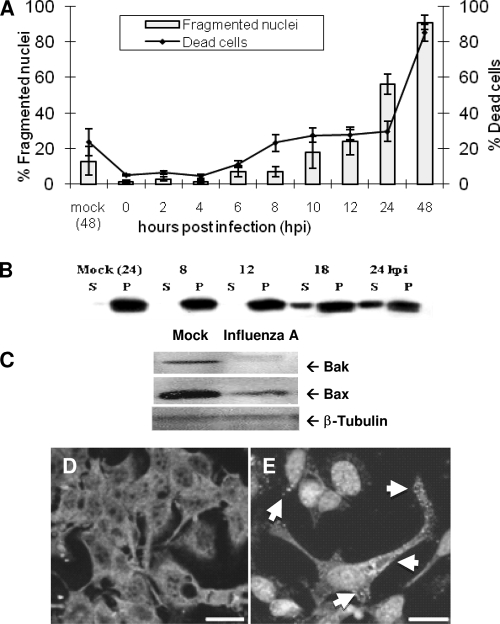
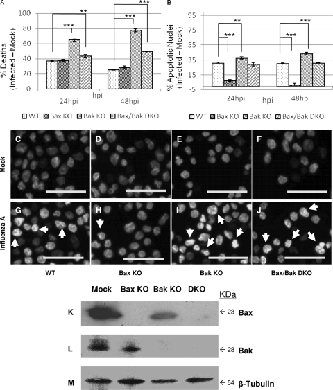
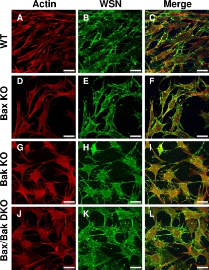
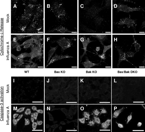
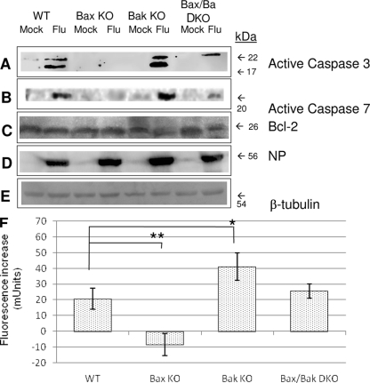
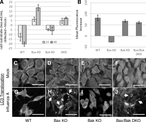
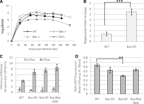
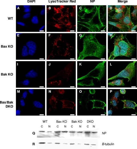
The ectopic overexpression of Bcl-2 restricts both influenza A virus-induced apoptosis and influenza A virus replication in MDCK cells, thus suggesting a role for Bcl-2 family members during infection. Here we report that influenza A virus cannot establish an apoptotic response without functional Bax, a downstream target of Bcl-2, and that both Bax and Bak are directly involved in influenza A virus replication and virus-induced cell death. Bak is substantially downregulated during influenza A virus infection in MDCK cells, and the knockout of Bak in mouse embryonic fibroblasts yields a dramatic rise in the rate of apoptotic death and a corresponding increase in levels of virus replication, suggesting that Bak suppresses both apoptosis and the replication of virus and that the virus suppresses Bak. Bax, however, is activated and translocates from the cytosol to the mitochondria; this activation is required for the efficient induction of apoptosis and virus replication. The knockout of Bax in mouse embryonic fibroblasts blocks the induction of apoptosis, restricts the infection-mediated activation of executioner caspases, and inhibits virus propagation. Bax knockout cells still die but by an alternative death pathway displaying characteristics of autophagy, similarly to our previous observation that influenza A virus infection in the presence of a pancaspase inhibitor leads to an increase in levels of autophagy. The knockout of Bax causes a retention of influenza A virus NP within the nucleus. We conclude that the cell and virus struggle to control apoptosis and autophagy, as appropriately timed apoptosis is important for the replication of influenza A virus.


Figures

FIG. 1.
FIG. 1.
Influenza A virus induces apoptosis through mitochondrial permeabilization via Bax activation. (A) Influenza A virus induction of MDCK cell death was assessed using trypan blue exclusion. Cell killing by influenza A/WSN/33 virus is pronounced by 24 hpi, with nearly total population death by 48 hpi (right axis, line). Nuclear alterations typical of apoptosis were observed by Hoechst staining and fluorescence microscopy. Dramatic nuclear fragmentation was seen by 24 hpi, and almost all cells exhibited nuclear fragmentation by 48 hpi (left axis, bars), indicating that influenza A virus-induced cell death is primarily apoptotic. (B) Cytochrome c release was assessed by centrifugal separation of mitochondria from the cytoplasm followed by Western blotting for cytochrome c. Cytochrome c was seen to shift from an exclusively mitochondrial localization (P) to the cytosolic compartment (S) by 18 hpi. (C) Bax and Bak expression in MDCK cells following infection was assessed by Western blotting. Bak, a protein that constitutively associates with the outer mitochondrial membrane, is severely downregulated following infection in MDCK cells. Bax is slightly downregulated by 24 hpi. β-Tubulin was used as a loading control. (D and E) Bax activation and translocation were assessed by immunocytochemistry and confocal microscopy. (D) Mock-infected cells show a diffuse staining pattern indicating inactive Bax located throughout the cytoplasm. (E) Upon activation by influenza A virus, Bax was seen to shift from its inactive cytoplasmic distribution to a punctate distribution (arrows) as activated Bax was translocated to the mitochondria following infection. In this figure, DAPI rather than Bax stains the nuclei. Bars, 50 μm.
FIG. 2.
FIG. 2.
Cell death and nuclear and mitochondrial alterations follow influenza A/WSN/33 virus infection in cells lacking Bax and/or Bak. (A) Total influenza virus-induced cell death was analyzed by trypan blue exclusion. While influenza A virus induces excessive cell death in Bak KO cells compared to the WT at 24 hpi and 48 hpi (P < 0.0001 and P < 0.0002, respectively), Bax KO cells do not differ from WT cells in the response to infection. Bax/Bak DKO cells show increased rates of death compared to the WT at 24 hpi and 48 hpi (P = 0.03 and P = 0.000001, respectively), indicating that Bax and Bak activities modulate death during infection. (B) Influenza A virus-induced nuclear condensation and fragmentation were analyzed by Hoechst staining. Bax KO cells exhibited a drastic reduction in nuclear alterations during influenza A virus infection compared to the WT at both 24 hpi and 48 hpi (P < 0.00003 and P = 0.00015, respectively). In contrast, Bak KO cells showed significant increases in influenza A virus-induced changes in nuclear morphology compared to the WT at 24 hpi and 48 hpi (P = 0.04 and P = 0.0002, respectively), while no significant difference was seen between Bax/Bak DKO and WT cells. These results suggest that influenza A virus may employ Bax as a means to trigger mitochondrial apoptosis during infection and that Bak antagonizes Bax in this situation. (C to J) Changes in nuclear morphology during influenza A virus infection were assessed by fluorescence microscopy at 48 hpi. Arrows mark nuclear condensation and fragmentation during influenza A virus infection, indicating apoptotic cell death. (C) WT mock; (D) WT plus influenza A virus; (E) Bax KO mock; (F) Bax KO plus influenza A virus; (G) Bak KO mock; (H) Bak KO plus influenza A virus; (I) Bax/Bak DKO mock; (J) Bax/Bak DKO plus influenza A virus. Bars, 50 μm. (K to M) KO of Bax and/or Bak was confirmed by Western blotting. (K and L) Bax is present only in WT and Bak KO cells (K), while Bak is present only in WT and Bax KO cells (L), confirming knockout in Bax KO, Bak KO, and Bax/Bak DKO cells. (M) β-Tubulin was used as a loading control.
FIG. 3.
FIG. 3.
Influenza A virus establishes infection and replicates in WT, Bax KO, Bak KO, and Bax/Bak DKO cells. Actin was stained by phalloidin-TRITC (A, D, G, and J). Immunocytochemical staining of influenza A virus-infected cells at 24 hpi with anti-WSN whole-virus primary antibody, followed by Alexa Fluor 488 staining (B, E, H, and K). Reveals cytoplasmic influenza A virus-induced vesicles containing mature virions in all infected cells regardless of Bax and/or Bak activity. (C, F, I, and L) Merge. Bars, 10 μm.
FIG. 4.
FIG. 4.
Influenza A virus-induced cytochrome c release and subsequent caspase activation are dependent primarily on Bax signaling. (A to H) Immunocytochemical staining of cytochrome c release in cells lacking Bax and/or Bak following influenza A virus infection. (A to D) Cytochrome c staining reveals a punctate distribution consistent with mitochondrial localization in healthy, mock-infected cells of each type. (E and G) Following influenza A virus infection at 24 hpi, cytoplasmic cytochrome c is evident in WT (E) and Bak KO (G) cells. (F and H) Bax KO (F) and Bax/Bak DKO (H) cells show no appreciable cytochrome c release by 48 hpi and retain a punctate cytochrome c staining pattern. These results indicate that Bax is necessary for influenza A virus-induced cytochrome c release. (I to P) Immunocytochemical staining of active caspase-3 in cells lacking Bax and/or Bak following influenza A infection. (I to L) Staining for active caspase-3 yields no signal in healthy, mock-infected cells. (M, O, and P) Following influenza A virus infection at 48 hpi, widespread caspase-3 activation is evident in WT (M), Bak KO (O), and Bax/Bak DKO (P) cells. (N) Bax KO cells do not show appreciable caspase-3 activation following influenza A virus infection, indicating that Bax, in the presence of Bak, is necessary for caspase-3 activation by influenza A virus. The activation of caspase-3 in Bax/Bak DKO cells indicates that there is an alternative pathway to caspase activation during influenza A virus infection in the absence of both Bax and Bak.
FIG. 5.
FIG. 5.
Executioner caspase activation does not occur after infection of Bax KO cells. (A to E) Western blotting for executioner caspase activation in influenza A virus-infected cells lacking Bax and/or Bak. Immunoblotting was performed with antibodies specific for active caspase-3 (A), cleaved caspase-7 (B), Bcl-2 (C), influenza A virus NP (D), and β-tubulin (E), which was used as a loading control. An activation of caspase-3 and caspase-7 was not seen in Bax KO MEFs and was impaired in Bax/Bak DKO cells, whereas caspase activation was evident in WT and Bak KO cells that constitutively expressed Bax. Bcl-2 expression was constant across all cell types in both mock- and influenza A virus-infected cells. (F) Executioner caspase activity (caspase-3 and caspase-7) was assessed using and Apo-One caspase activity kit, and cleavage of fluorogenic substrate was assessed at 495 nm by use of a fluorescence microplate reader (Bio-Tek). Data are expressed as arbitrary units (milliunits [mUnits]), (fluorescence from influenza A virus-infected cells) − (fluorescence from mock-infected cells), in individual wells. The rate of activated executioner caspase substrate cleavage was high in Bak KO cells compared to the WT (P = 0.03), but the substrate was largely uncleaved in Bax KO cells following infection (P = 0.007), indicating that Bax in the presence of Bak is required for executioner caspase activation in response to influenza A virus.
FIG. 6.
FIG. 6.
Influenza A virus induces autophagy-like death in the absence of Bax. (A) Lysosomal acid phosphatase activity in influenza A virus-infected cells was assessed by measuring the release of p-nitrophenol from p-nitrophenylphosphate substrate. In cells constitutively expressing Bax, acid phosphatase activity decreases following infection, as these cells follow an apoptotic path to death. In cells lacking Bax, acid phosphatase activity increased, indicating an increase in lysosomal activity, a common marker for autophagic death. One unit equals 1 μmol of 4-nitrophenylphosphate per minute under the experimental conditions. (B) Cells were infected for 48 hpi and stained with Lysotracker Red DND-99 for 30 min prior to collection and immediate analysis by FACS. In WT, Bak KO, and Bax/Bak DKO cells, influenza A virus infection results in an increase in lysosomal volume by 48 hpi. In Bax KO cells, a slight decrease in lysosomal volume was observed by 48 hpi. (C to J) Cells were transfected with a construct expressing LC3-GFP prior to infection and observed by confocal microscopy for LC3 expression and translocation following infection. (C to F) The diffuse LC3-GFP expression pattern in mock-infected cells indicates a cytoplasmic, inactive distribution of LC3 in each cell type. (G and I) In cells constitutively expressing Bax, LC3-GFP expression remains diffuse following infection, indicating a lack of LC3 activation as these cells undergo apoptosis. (H and J) In cells lacking Bax (Bax KO and Bax/Bak DKO cells), LC3-GFP staining shifts to a punctate pattern, indicating LC3 activation and translocation to autophagosomes, a process that occurs solely during autophagy. Bars, 25 μm.
FIG. 7.
FIG. 7.
Influenza A virus replication is dependent upon opposite virus-induced effects on Bax and Bak activity that are unlikely to be interferon related. (A) Influenza A virus replication was analyzed by plaque assay. Virus replication is severely attenuated in Bax KO cells, resulting in a 2-log decrease in PFU/ml compared to the WT. Bak KO cells allow a maximum replication similar to that of the WT, while Bax/Bak DKO cells show a slight elevation of infectious titers during infection. These results indicate that Bax is proviral during infection, while Bak is dispensable for replication. (B) Bax was transiently expressed in all cell types by Lipofectamine 2000 transfection of a C2-Bax-GFP construct prior to infection, and supernatant samples were collected for plaque assay at 48 hpi. Baseline virus replication in each cell type was evaluated using empty C2-GFP plasmid transfection. Bax reconstitution in Bax KO cells resulted in a fivefold increase in infectious titers compared to the control (P = 0.0007). A minimal effect on the virus titer was seen after Bax overexpression by transient transfection in WT cells compared to empty plasmid controls. (C) Influenza A virus replication was assessed by reverse transcription-PCR. Serial dilutions of stock virus at known concentrations were also analyzed to generate a standard curve to which experimental samples were compared, thus calculating the approximate number of influenza A virus particles/ml in each sample. By 24 hpi, Bax KO, Bak KO, and Bax/Bak DKO cells all showed significantly higher levels of influenza A virus RNA released into the cell culture supernatant than did WT cells. (D) Interferon activity was assessed by infecting mock- and influenza A virus-infected cells with interferon-sensitive, GFP-linked NDV and quantifying the mean GFP expression levels of 10,000 events per condition by FACS analysis. Each assay was run in triplicate, and data are expressed as the ratio of the numbers of influenza A virus-infected to mock-infected cells per cell type. After influenza A virus infection, Bak KO cells exhibited a slight decrease in ratio compared to the WT, representing a 30% increase in interferon activity (P = 0.002). Bax KO and Bax/Bak DKO cells both showed similar fluorescence changes compared to the WT after infection. Due the high degree of similarity between cell types, these results suggest that the interferon response in infected cells is modulated by viral replication in the presence of Bak and is only slightly modified by Bax activity during influenza A virus infection. As an elevated interferon response typically leads to a reduced virus replication capacity, these results also suggest that it is unlikely that the observed trends in infectious virus titer are due to virus-induced interferon signaling.
FIG. 8.
FIG. 8.
Influenza A virus NP exhibits increased nuclear retention in Bax KO cells. Nuclear retention of NP due to a lack of caspase activity has been linked to decreased titers of virus. DAPI staining was used for nuclear localization (A, E, I, and M), lysosomes were stained with LysoTracker Red DND-99 (B, F, J, and N), and influenza A virus NP localization was determined using an NP-specific antibody (C, G, K, and O). Images were taken using a confocal microscope. (D, H, L, and P) Merge. Lysosomal localization of influenza A virus NP was not observed in WT cells or any of the KO cells. NP showed near-complete cytoplasmic localization in WT cells (D) and Bax/Bak DKO cells (P). A slight nuclear retention of NP was observed in Bak KO cells (L), while in Bax KO cells (H), a nuclear retention of influenza A virus NP is obvious in all infected cells. Bars, 10 μm. (Q) Nuclear retention of NP in infected Bax KO and, to a lesser extent, Bak KO cells was confirmed by Western blotting of cytoplasmic (C) and nuclear (N) fractions of infected cells. (R) β-Tubulin was used to ensure efficient fractionation.

Similar articles

Cited by

References

    1. Bergmann, M., A. Garcia-Sastre, E. Carnero, H. Pehamberger, K. Wolff, P. Palese, and T. Muster. 2000. Influenza virus NS1 protein counteracts PKR-mediated inhibition of replication. J. Virol. 746203-6206. - PMC - PubMed
    1. Boya, P., K. Andreau, D. Poncet, N. Zamzami, J.-L. Perfettini, D. Metivier, D. M. Ojcius, M. Jaattela, and G. Kroemer. 2003. Lysosomal membrane permeabilization induces cell death in a mitochondrion-dependent fashion. J. Exp. Med. 1971323-1334. - PMC - PubMed
    1. Brydon, E. W. A., S. J. Morris, and C. Sweet. 2005. Role of apoptosis and cytokines in influenza virus morbidity. FEMS Microbiol. Rev. 29837-850. - PubMed
    1. Brydon, E. W. A., H. Smith, and C. Sweet. 2003. Influenza A virus-induced apoptosis in bronchiolar epithelial (NCI-H292) cells limits pro-inflammatory cytokine release. Soc. Gen. Microbiol. 842389-2400. - PubMed
    1. Chen, W., P. A. Calvo, and D. Malide. 2001. A novel influenza A virus mitochondrial protein that induces cell death. Nat. Med. 71306-1312. - PubMed

Publication types