. 2009 Jul 24;284(30):20197-205.
doi: 10.1074/jbc.M109.026096. Epub 2009 Jun 3.

Acetylation of the DNA binding domain regulates transcription-independent apoptosis by p53

Affiliations

Acetylation of the DNA binding domain regulates transcription-independent apoptosis by p53

Stephen M Sykes et al. J Biol Chem. .

Abstract

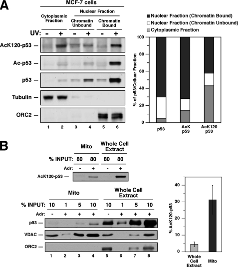
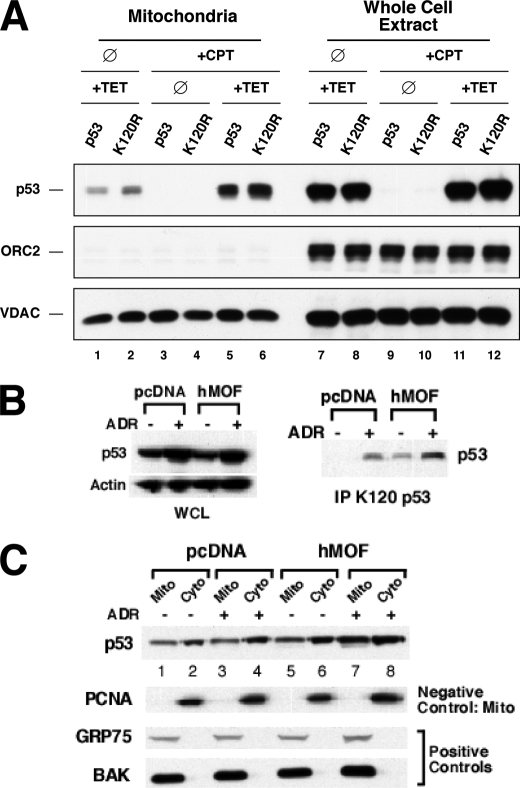
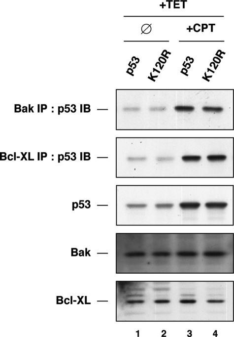
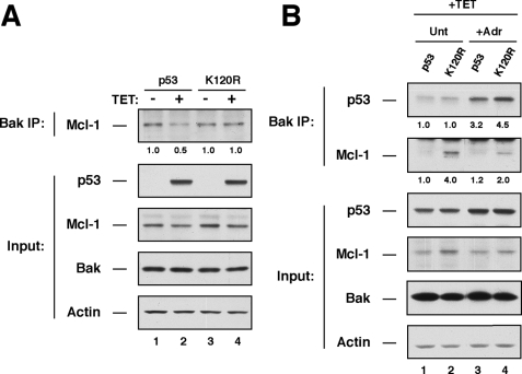
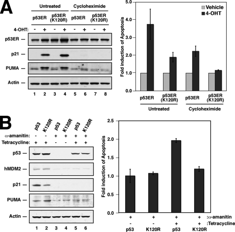
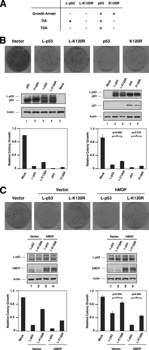
The tumor suppressor p53 induces apoptosis by altering the transcription of pro-apoptotic targets in the nucleus and by a direct, nontranscriptional role at the mitochondria. Although the post-translational modifications regulating nuclear apoptotic functions of p53 have been thoroughly characterized, little is known of how transcription-independent functions are controlled. We and others identified acetylation of the p53 DNA binding domain at lysine 120 as a critical event in apoptosis induction. Although initial studies showed that Lys-120 acetylation plays a role in p53 function in the nucleus, we report here a role for Lys-120 acetylation in transcription-independent apoptosis. We demonstrate that the Lys-120-acetylated isoform of p53 is enriched at mitochondria. The acetylation of Lys-120 does not appear to regulate the ability of p53 to interact with the pro-apoptotic proteins BCL-XL and BAK. However, displacement of the inhibitory MCL-1 protein from BAK is compromised when Lys-120 acetylation is blocked. Functional studies show that mutation of Lys-120 to a nonacetylated residue, as occurs in human cancer, inhibits transcription-independent apoptosis, and enforced acetylation of Lys-120 enhances transcription-independent apoptosis. These data support a model whereby Lys-120 acetylation contributes to both the transcription-dependent and -independent apoptotic pathways induced by p53.


Figures

FIGURE 1.
FIGURE 1.
Acetylated Lys-120-p53 is enriched at chromatin, cytoplasmic, and mitochondrial fractions. A, 18 h following 7.5 J/m2 UV exposure, MCF-7 cells were harvested and biochemically fractionated into nuclear fraction (chromatin-bound and -unbound) and cytoplasmic fractions. Subsequently, each fraction was divided into equal aliquots and subjected to immunoprecipitation with antibodies that recognize either any acetylated protein or specifically the p53 acetylated at lysine 120 (AcK120-p53). Precipitates were then analyzed by Western blot with p53-specific antibodies. Input lysates were also blotted with p53, tubulin (cytoplasmic marker), and ORC2 (nuclear marker) antibodies. B, U2OS cells treated with 0.5 μm adriamycin (Adr) for 18 h and untreated U2OS cells were harvested and separated into 2 equal aliquots. One aliquot from each condition was lysed with a CHAPS-based lysis buffer (described under “Experimental Procedures”) to generate whole cell extracts. Cells from the 2nd aliquot were biochemically fractionated to isolate mitochondria (Mito) and subsequently lysed with a CHAPS-based lysis buffer. Following lysis, 80% of mitochondrial and whole cell extracts were subjected to immunoprecipitation with the AcLys-120 antibody. Precipitates were subjected to Western blot with p53 antibodies to quantify p53 acetylated at lysine 120 (top panel). The amount of p53 localized to the mitochondria is relatively small compared with the amount of total p53 in a stressed cell. Therefore, to compare p53 protein levels between the mitochondria and total p53 lysates in the linear range, 1, 5, and 10% of input lysate from each adriamycin-treated mitochondrial extract (bottom 3 panels, lanes 2–4, respectively) and whole cell extracts (bottom 3 panels, lanes 6–8, respectively) were blotted with p53, voltage-dependent anion-selective channel (mitochondrial marker), and ORC2 (nuclear marker) antibodies. Furthermore, 10% of each untreated fraction was also blotted with the antibodies described (lanes 1 and 5). The acetyl-Lys-120 signals (top panel) from each sample were compared with the titrations probed for total p53 (lower panel). The bar graph to the right represents the percentage of p53 in whole cell extracts acetylated at lysine 120 (light gray, 5 ± 1%) versus the percentage of mitochondrial p53 acetylated at lysine 120 (dark gray, 32 ± 8%).
FIGURE 2.
FIGURE 2.
Lys-120 acetylation is dispensable for p53 mitochondrial localization. A, H1299 cells expressing either p53 or the K120R mutant under the regulation of a tetracycline (TET)-response element were treated with 500 ng/ml tetracycline or vehicle for 6 h. Following tetracycline treatment, cells were administered 5 μm camptothecin (CPT) or vehicle for an additional 18 h. After treatment, cells were harvested and biochemically fractionated into mitochondria or whole cell extracts. Subsequently, each fraction was subjected to Western blot analysis with p53, voltage-dependent anion-selective channel (mitochondrial marker), and ORC2 (nuclear marker) antibodies. ∅, vehicle-treated. B and C, H1299 cells expressing p53 under the regulation of a tetracycline-response element were transfected with either hMOF or empty vector. Following transfection, cells were treated with 500 ng/ml tetracycline 6 h and then cells were administered 0.5 μm adriamycin (ADR) or vehicle for an additional 18 h. After treatment, cells were harvested and biochemically fractionated into mitochondria (Mito), cytoplasmic (Cyto), and whole cell extracts (WCE). Subsequently, each fraction was subjected to Western blot analysis with the indicated antibodies. PCNA, proliferating cell nuclear antigen.
FIGURE 3.
FIGURE 3.
Lys-120 acetylation is dispensable for p53 to interact with BAK or BCL-XL. H1299 cells expressing either p53 or the K120R mutant under the regulation of a tetracycline (TET)-response element were treated with 500 ng/ml tetracycline or vehicle for 6 h. Following tetracycline treatment, cells were administered 5 μm camptothecin (CPT) or vehicle for an additional 18 h. After treatment, cells were collected and fractionated in nuclear and cytoplasmic fractions. Cytoplasmic fractions were subjected to immunoprecipitation with either BAK or BCL-XL antibodies. Precipitates were then analyzed by Western blot with p53 antibodies. Input lysates were also blotted with p53, BAK, and BCL-XL antibodies. IB, immunoblot; ∅, vehicle-treated.
FIGURE 4.
FIGURE 4.
Lys-120 acetylation is required for p53 to displace the anti-apoptotic protein MCL-1 from BAK. A, H1299 cells expressing either p53 or the K120R mutant under the regulation of a tetracycline (TET)-response element were treated with 500 ng/ml tetracycline or vehicle for 24 h. After treatment, cells were collected and lysed to generate whole cell extracts. Whole cell extracts were the subjected to immunoprecipitation with BAK antibodies. BAK precipitates were then analyzed by Western blot with antibodies that recognize p53 or MCL-1. Input lysates were also blotted with p53, BAK, MCL-1, and actin antibodies. B, H1299 cells expressing either p53 or the K120R mutant under the regulation of a tetracycline-response element were treated with 500 ng/ml tetracycline or vehicle for 6 h. Following tetracycline treatment, cells were administered 0.5 μm adriamycin (Adr) or vehicle for an additional 18 h. After treatment, cells were collected and analyzed as in A. Unt, untreated.
FIGURE 5.
FIGURE 5.
Annexin V staining demonstrates that lysine 120 acetylation is required for p53-mediated transcription-independent apoptosis. A, H1299 cells expressing either p53ER or the p53ER(K120R) mutant were treated with 100 μg/ml cycloheximide or vehicle for 1 h and subsequently treated with 100 nm 4-OHT or vehicle for an additional 48 h. Following treatment, cells were collected and either subjected to Western blot analysis with the indicated antibodies (left panel) or stained with annexin V-FITC and propidium iodide and then analyzed by fluorescence-activated cell sorter (right panel). Right panel is a graphic representation of annexin V-positive (propidium iodide-negative) cells collected after the treatment indicated. Apoptosis levels were normalized to “fold induction” as discussed in the text, and basal levels of apoptosis are ∼2% in the absence of CHX treatment and 15% in the presence of CHX. Error bars represent standard deviation of three independent reactions. B, H1299 cells expressing either p53 or the K120R mutant under the regulation of a tetracycline-response element were treated with 100 μg/ml α-amanitin or vehicle for 2 h and subsequently administered 500 ng/ml tetracycline or vehicle for an additional 24 h. Following treatment, cells were collected and either subjected to Western blot analysis with the indicated antibodies (left panel) or stained with annexin V-FITC and propidium iodide and then analyzed by fluorescence-activated cell sorter (right panel). Right panel is a graphical representation of annexin V-positive (propidium iodide-negative) cells collected after the treatment indicated. Levels of apoptosis are expressed as fold induction. Basal levels of apoptosis were ∼5% in the absence of tetracycline, which is similar to the level observed in response to α-amanitin treatment of parental H1299 cells. Upon activation of p53 by tetracycline treatment, the number of apoptotic cells reached ∼10% of the total population. Error bars represent standard deviation of three independent reactions.
FIGURE 6.
FIGURE 6.
Colony growth assays demonstrates that lysine 120 acetylation is required for the transcription-independent functions of p53. A, H1299 cells were transiently transfected with plasmids expressing either p53, K120R, or each fused with a mitochondrial import signal (L-p53 and L-K120R). Nontagged versions of p53 are capable of inducing cell cycle arrest, and transcription-dependent (TDA) and -independent apoptosis (TIA). In contrast, the L-p53 isoforms are competent exclusively for transcription-independent apoptosis, as indicated. B, following transfection of the wild-type and mitochondria-targeted p53 isoforms, cells were replated at low density and cultured in media supplemented with G418 for 12 days, then fixed, and stained. Plates of stained cells from experiment 1 are shown (top panel) and colony numbers quantified (left graph). This analysis was repeated, and the quantitation of colony numbers for experiment 2 is displayed (right graph). Western blotting documented appropriate expression levels of p53 isoforms in experiments 1 and 2, as indicated. Experiment 2 was performed as biological triplicates, and results are displayed with standard errors indicated. In addition, p values were generated (Student's t test) by comparing the values for the wild type and K120R isoforms of p53, in the nontagged (p ≤ 0.018) and mitochondria-tagged isoforms (p ≤ 0.006). C, H1299 cells were transfected with the mitochondria-targeted L-p53 constructs described in A, along with an hMOF expression vector where indicated. Colony formation was assayed 12 days post-transfection as described above. Stained colonies from experiment 1 are displayed (top panel) and quantitated (left graph). In experiment 2, biological triplicates were performed, and colony number was quantified (right graph). Western blotting documented appropriate expression levels of p53 isoforms and hMOF in experiments 1 and 2, as indicated. For experiment 2, standard error bars are displayed. In addition, p values were generated (Student's t test) by comparing the values for the wild type and K120R isoforms of p53, in the presence (p ≤ 0.003) and absence of hMOF (p ≤ 0.032).

Similar articles

Cited by

References

    1. Vousden K. H., Lu X. (2002) Nat. Rev. Cancer 2, 594–604 - PubMed
    1. el-Deiry W. S., Tokino T., Velculescu V. E., Levy D. B., Parsons R., Trent J. M., Lin D., Mercer W. E., Kinzler K. W., Vogelstein B. (1993) Cell 75, 817–825 - PubMed
    1. Chipuk J. E., Green D. R. (2006) Cell Death Differ. 13, 994–1002 - PubMed
    1. Kuribayashi K., El-Deiry W. S. (2008) Adv. Exp. Med. Biol. 615, 201–221 - PubMed
    1. Erster S., Mihara M., Kim R. H., Petrenko O., Moll U. M. (2004) Mol. Cell. Biol. 24, 6728–6741 - PMC - PubMed

Publication types

MeSH terms

LinkOut - more resources