. 2009 Jun 3;29(22):7124-36.
doi: 10.1523/JNEUROSCI.1090-09.2009.

PSD-95 is essential for hallucinogen and atypical antipsychotic drug actions at serotonin receptors

Affiliations

PSD-95 is essential for hallucinogen and atypical antipsychotic drug actions at serotonin receptors

Atheir I Abbas et al. J Neurosci. .

Abstract

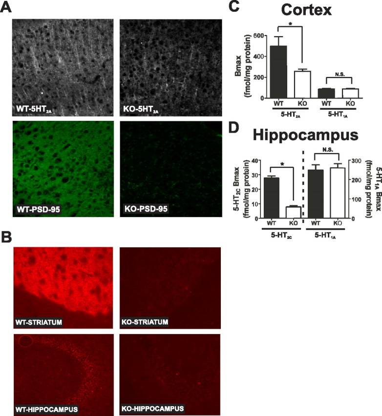
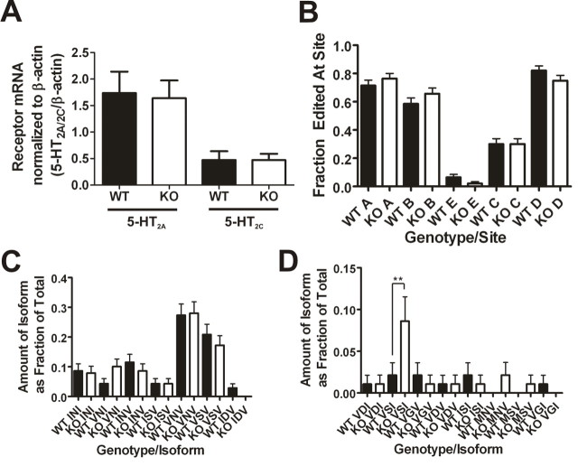
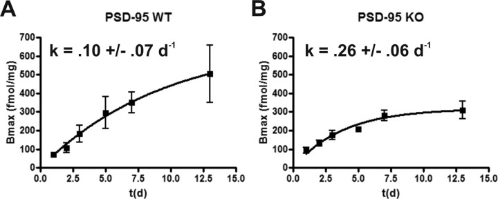
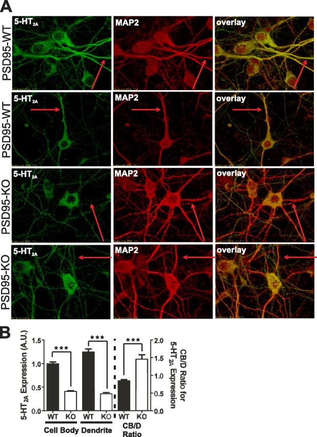
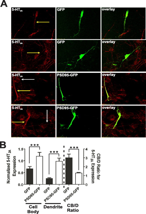
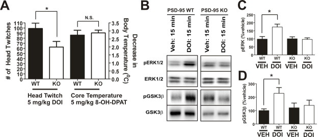
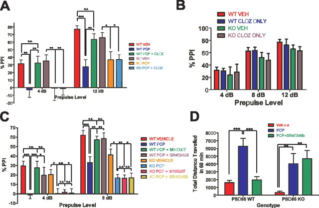
Here, we report that postsynaptic density protein of 95 kDa (PSD-95), a postsynaptic density scaffolding protein, classically conceptualized as being essential for the regulation of ionotropic glutamatergic signaling at the postsynaptic membrane, plays an unanticipated and essential role in mediating the actions of hallucinogens and atypical antipsychotic drugs at 5-HT(2A) and 5-HT(2C) serotonergic G-protein-coupled receptors. We show that PSD-95 is crucial for normal 5-HT(2A) and 5-HT(2C) expression in vivo and that PSD-95 maintains normal receptor expression by promoting apical dendritic targeting and stabilizing receptor turnover in vivo. Significantly, 5-HT(2A)- and 5-HT(2C)-mediated downstream signaling is impaired in PSD-95(null) mice, and the 5-HT(2A)-mediated head-twitch response is abnormal. Furthermore, the ability of 5-HT(2A) inverse agonists to normalize behavioral changes induced by glutamate receptor antagonists is abolished in the absence of PSD-95 in vivo. These results demonstrate that PSD-95, in addition to the well known role it plays in scaffolding macromolecular glutamatergic signaling complexes, profoundly modulates metabotropic 5-HT(2A) and 5-HT(2C) receptor function.


Figures

Figure 1.
Figure 1.
Genetic deletion of PSD-95 results in a selective loss of 5-HT2A and 5-HT2C receptors. A, 5-HT2A and PSD-95 double-label immunochemistry in medial prefrontal cortex of PSD-95wildtype and PSD-95null mice shows a large reduction in 5-HT2A receptor expression in null mice (N = 3 littermate pairs). B, 5-HT2C immunochemistry in PSD-95wildtype and PSD-95null striatum and hippocampus reveals that 5-HT2C receptor expression is almost completely abolished in the absence of PSD-95 in both striatum and hippocampus (N = 3 littermate pairs). C, Comparison of B max estimates for the 5-HT2A receptor (N = 4 littermate pairs) and the 5-HT1A receptor (N = 5 littermate pairs) in PSD-95wildtype and PSD-95null cortices. B max estimates were obtained by performing [3H]-ketanserin (5-HT2A) and [3H]-WAY100635 (5-HT1A) saturation binding on microdissected and homogenized cortical tissue. Quantitation showed an ∼40% reduction in 5-HT2A expression and no change in 5-HT1A expression in the cortices of PSD-95null mice. D, Comparison of B max estimates for the 5-HT2C receptor (N = 3; tissue from 3 animals was pooled for each measurement, for a total of 9 animals, all littermate pairs) and 5-HT1A receptor (N = 6 littermate pairs) in PSD-95wildtype and PSD-95null hippocampi. B max estimates were obtained by performing [3H]mesulergine saturation binding in the presence of 100 nm spiperone to block the vast majority of 5-HT2A receptors (5-HT2C) and [3H]-WAY100635 saturation binding (5-HT1A). Quantitation showed an almost 70% reduction in 5-HT2C expression and no change in 5-HT1A expression in hippocampus in the absence of PSD-95. All saturation binding was analyzed using nonlinear least squares fitting. B max data are presented as means ± SEM; *p < 0.05, **p < 0.01, ***p < 0.001; one-tailed unpaired t test.
Figure 2.
Figure 2.
Genetic deletion of PSD-95 does not affect mRNA levels of 5-HT2A and 5-HT2C receptors or mRNA editing of the 5-HT2C receptor. A, Cortical 5-HT2A receptor mRNA levels (N = 4 littermate pairs; 11 measurements were performed for each animal) and hippocampal 5-HT2C receptor mRNA levels (N = 4 littermate pairs; 5 measurements for each animal) normalized to β-actin mRNA levels as measured by quantitative RT-PCR. There are no changes in 5-HT2A mRNA levels and no changes in 5-HT2C mRNA levels in the absence of PSD-95. B, 5-HT2C mRNA editing frequencies at five edited sites. The frequency of editing at the five sites is not significantly different in PSD-95null mice. C, D, Frequencies of the different edited isoforms detected (wildtypes, N = 94; nulls, N = 93). Fifteen isoforms were detected, and 14 of them were not significantly altered in the absence of PSD-95. There is a significant increase in the proportion of the VSI isoform in PSD-95null mouse hippocampus. All mRNA editing data are plotted as the frequency expressed as a fraction of the total, ± the SEM. Normalized mRNA measurements are presented as means ± SEM; *p < 0.05, **p < 0.01, ***p < 0.001; one-tailed unpaired t test for the comparison of mRNA levels and one-way ANOVA followed by Newman–Keuls post hoc tests for comparison of the mRNA editing measurements.
Figure 3.
Figure 3.
Genetic deletion of PSD-95 leads to an accelerated turnover of 5-HT2A receptor protein. A and B represent fitted curves modeling 5-HT2A receptor turnover in PSD-95wildtype and PSD-95null mice, respectively (N = 3–4 littermate pairs at each data point). Visual inspection shows that steady-state levels for the 5-HT2A receptor are reached sooner in the absence of PSD-95, suggesting accelerated turnover. The higher k in PSD-95null cortex indicates a higher rate of receptor turnover in the absence of PSD-95. Rate constant, k, is a nonleast squares fitted parameter of an equation modeling receptor recovery (for details, see Materials and Methods), ± SEM; *p < 0.05, **p < 0.01, ***p < 0.001; one-tailed unpaired t test.
Figure 4.
Figure 4.
5-HT2A receptor expression and dendritic targeting is attenuated in neurons prepared from PSD-95null mice. A, Representative images of double-label immunochemistry performed on P0.5 cortical neurons of PSD-95wildtype and PSD-95null mice. The red arrows highlight the same dendritic process in all three images of each neuron. MAP2-positive dendrites display reduced 5-HT2A receptor expression in the absence of PSD-95. B, Comparison of 5-HT2A receptor expression, normalized to MAP2 expression, in cell bodies and dendrites, and CB/D expression ratio, in PSD-95wildtype and PSD-95null neurons. Quantitation of 5-HT2A receptors shows that in PSD-95null neurons, expression is dramatically reduced in both neuronal soma and dendrites. The increase in the CB/D ratio in PSD-95null neurons suggests an impairment in dendritic targeting in the absence of PSD-95. N = 3 littermate pairs, 17 neurons from each animal, for a total of 51 neurons measured per genotype. Data are presented as the mean ± the SEM; *p < 0.05, **p < 0.01, ***p < 0.001; one-tailed unpaired t test.
Figure 5.
Figure 5.
Reintroduction of PSD-95 rescues 5-HT2A receptor expression and trafficking deficits in PSD-95null cortical neurons in vitro. A, Representative images of double-label immunochemistry performed on P0.5 cortical neurons of PSD-95null mice infected with either GFP lentivirus (top 2 rows of panels) or PSD-95–GFP lentivirus (bottom 2 rows of panels). PSD-95null neurons from each animal were plated in two wells, one for GFP lentiviral infection and the other for PSD-95–GFP lentiviral infection. The yellow arrows highlight dendritic 5-HT2A receptor expression in an infected neuron. White arrows highlight 5-HT2A receptor expression in an uninfected neuron. GFP-infected neurons display low overall 5-HT2A expression and low dendritic targeting. In contrast, PSD-95–GFP-infected neurons display a dramatic increase in overall 5-HT2A receptor expression and substantially more receptor appears to be targeted to the dendritic compartment, both compared with control GFP-infected neurons and compared with uninfected neurons in the same field. B, Comparison of 5-HT2A receptor expression in cell bodies and dendrites and the CB/D ratio in GFP- and PSD-95–GFP-infected PSD-95null neurons. Expression is normalized to GFP or PSD-95–GFP. PSD-95–GFP leads to substantial rescue of 5-HT2A receptor expression compared with GFP-infected control neurons. PSD-95–GFP addback leads to a significant reduction in the CB/D ratio compared with GFP-expressing control neurons, suggesting that PSD-95–GFP is rescuing dendritic targeting of 5-HT2A receptor expression. N = 3 animals for each animal/lentivirus, and 10 infected neurons from each animal/lentivirus were measured (60 neurons total). Data are presented as the mean ± the SEM; *p < 0.05, **p < 0.01, ***p < 0.001; one-tailed unpaired t test.
Figure 6.
Figure 6.
Deletion of PSD-95 attenuates 5-HT2C receptor-mediated induction of c-fos. A, 5-HT2C and c-fos double-label immunochemistry in the hippocampus of PSD-95wildtype and PSD-95null mice after MK-212 treatment (N = 3 littermate pairs). Representative images of CA1 and CA2 are shown. There are fewer c-fos-positive cells in the PSD-95null mice treated with MK-212 in both CA1 and CA2. B, Higher magnification image of CA1 to examine colocalization of 5-HT2C receptors and c-fos. 5-HT2C receptor colocalizes with c-fos, suggesting that 5-HT2C is inducing this IEG directly, rather than indirectly in surrounding neurons. C, Analysis of c-fos induction was performed by counting the number of c-fos-positive cells in CA1, CA2, CA3, and DG. Data are presented as the mean number of c-fos-positive cells ± the SEM. There is a significant reduction in c-fos-positive cells in the absence of PSD-95 in all four regions that were measured. c-fos counts were performed separately in the hippocampus of each hemisphere (2 values for each section analyzed). Every seventh section was analyzed, for a total of six sections per animal. *p < 0.05, **p < 0.01, ***p < 0.001; one-tailed unpaired t test.
Figure 7.
Figure 7.
5-HT2A-mediated head-twitch behavior is attenuated in PSD-95null mice. A, 5-HT2A-mediated head-twitch response after intraperitoneal injection of 5 mg/kg DOI (N = 11 littermate pairs) and 5-HT1A-mediated hypothermic response after intraperitoneal injection of 5 mg/kg 8-OH-DPAT (N = 12 littermate pairs). There is a significant decrease in DOI-induced head-twitch in PSD-95null mice, in contrast with the magnitude of the decrease in core body temperature induced by 8-OH-DPAT, which is the same in both PSD-95wildtype and PSD-95null mice. B–D, Western blot and associated quantification for p-ERK1/2, ERK1/2, p-GSK3β, and GSK3β 15 min after intraperitoneal injection of vehicle or DOI (N = 4–5 littermate pairs). DOI induces pERK1/2 and pGSK3β in wild-type, but not null, mice. For quantification, p-ERK1/2 and p-GSK3β were normalized to ERK1/2 and GSK3β, respectively. Data are given as means ± the SEM; *p < 0.05, **p < 0.01, ***p < 0.001; one-tailed unpaired t test.
Figure 8.
Figure 8.
The antipsychotic-like efficacy of atypical antipsychotics is lost in PSD-95null mice. At the two prepulses shown, 4 and 12 dB, PCP significantly disrupted PPI in PSD-95wildtype and PSD-95null mice. In PSD-95wildtype mice, clozapine pretreatment normalized the disruption of PPI by PCP at both 4 and 12 dB (N = 6 littermate pairs). In contrast, in PSD-95null mice, clozapine had no antipsychotic-like effect. B, PPI after treatment with clozapine alone (N = 6 littermate pairs). Clozapine (0.5 mg/kg) had no effect on PPI compared with vehicle. C, PPI in PSD-95wildtype and PSD-95null mice after injection of vehicle, 6.0 mg/kg PCP, 0.5 mg/kg M100907 plus 6.0 mg/kg PCP, or 1.0 mg/kg SR46349B plus 6.0 mg/kg PCP. PCP significantly disrupted PPI in PSD-95wildtype and PSD-95null mice at 4 and 8 dB (N = 12). In PSD-95wildtype mice, M100907 or SR46349B pretreatment normalized the disruption of PPI by PCP at 4 and 8 dB, whereas in PSD-95null mice, they had no antipsychotic-like effect. D, Locomotion after vehicle, 6.0 mg/kg PCP, and 1.0 mg/kg SR46349B plus 6.0 mg/kg PCP in PSD-95wildtype and PSD-95null mice (N = 12). SR46349B normalizes PCP-induced hyperlocomotion in PSD-95wildtype mice only. Data are given as means ± the SEM; *p < 0.05, **p < 0.01, ***p < 0.001; two-way repeated-measures ANOVA followed by Bonferroni post-tests.

References

    1. Aghajanian GK, Marek GJ. Serotonin and hallucinogens. Neuropsychopharmacology. 1999;21:16S–23S. - PubMed
    1. Ahlemeyer B, Baumgart-Vogt E. Optimized protocols for the simultaneous preparation of primary neuronal cultures of the neocortex, hippocampus and cerebellum from individual newborn (P0.5) C57BL/6J mice. J Neurosci Methods. 2005;149:110–120. - PubMed
    1. Beaulieu JM, Marion S, Rodriguiz RM, Medvedev IO, Sotnikova TD, Ghisi V, Wetsel WC, Lefkowitz RJ, Gainetdinov RR, Caron MG. A beta-arrestin 2 signaling complex mediates lithium action on behavior. Cell. 2008;132:125–136. - PubMed
    1. Bécamel C, Alonso G, Galéotti N, Demey E, Jouin P, Ullmer C, Dumuis A, Bockaert J, Marin P. Synaptic multiprotein complexes associated with 5-HT(2C) receptors: a proteomic approach. EMBO J. 2002;21:2332–2342. - PMC - PubMed
    1. Bécamel C, Gavarini S, Chanrion B, Alonso G, Galéotti N, Dumuis A, Bockaert J, Marin P. The serotonin 5-HT2A and 5-HT2C receptors interact with specific sets of PDZ proteins. J Biol Chem. 2004;279:20257–20266. - PubMed

Publication types

MeSH terms

Substances