

Corticocortical interactions between and within three cortical auditory areas specialized for time-domain signal processing
- PMID: 19494145
- PMCID: PMC2752974
- DOI: 10.1523/JNEUROSCI.0373-09.2009
Corticocortical interactions between and within three cortical auditory areas specialized for time-domain signal processing
Abstract
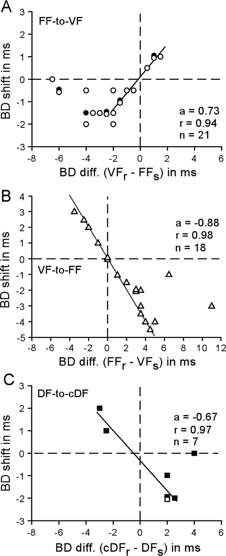
In auditory cortex of the mustached bat, the FF (F means frequency modulation), dorsal fringe (DF), and ventral fringe (VF) areas consist of "combination-sensitive" neurons tuned to the pair of an emitted biosonar pulse and its echo with a specific delay (best delay: BD). The DF and VF areas are hierarchically at a higher level than the FF area. Focal electric stimulation of the FF area evokes "centrifugal" BD shifts of DF neurons, i.e., shifts away from the BD of the stimulated FF neurons, whereas stimulation of the DF neurons evokes "centripetal" BD shifts of FF neurons, i.e., shifts toward the BD of the stimulated DF neurons. In our current studies, we found that the feedforward projection from FF neurons evokes centrifugal BD shifts of VF neurons, that the feedback projection from VF neurons evokes centripetal BD shifts of FF neurons, that the contralateral projection from DF neurons evokes centripetal BD shifts of DF neurons, and that the centripetal BD shifts evoked by the DF and VF neurons are 2.5 times larger than the centrifugal BD shifts evoked by the FF neurons. The centrifugal BD shifts shape the selective neural representation of a specific target distance, whereas the centripetal BD shifts expand the representation of the selected specific target distance to focus on the processing of the target information at a specific distance. The centrifugal and centripetal BD shifts evoked by the feedforward and feedback projections promote finer analysis of a target at shorter distances.
Figures


Similar articles
-
Modulation of auditory processing by cortico-cortical feed-forward and feedback projections.Proc Natl Acad Sci U S A. 2008 May 27;105(21):7600-5. doi: 10.1073/pnas.0802961105. Epub 2008 May 21. Proc Natl Acad Sci U S A. 2008. PMID: 18495931 Free PMC article.
-
Reorganization of the cochleotopic map in the bat's auditory system by inhibition.Proc Natl Acad Sci U S A. 2002 Nov 26;99(24):15743-8. doi: 10.1073/pnas.242606699. Epub 2002 Nov 5. Proc Natl Acad Sci U S A. 2002. PMID: 12419852 Free PMC article.
-
Bilateral cortical interaction: modulation of delay-tuned neurons in the contralateral auditory cortex.J Neurosci. 2007 Aug 1;27(31):8405-13. doi: 10.1523/JNEUROSCI.1257-07.2007. J Neurosci. 2007. PMID: 17670987 Free PMC article.
-
Plasticity of the adult auditory system based on corticocortical and corticofugal modulations.Neurosci Biobehav Rev. 2020 Jun;113:461-478. doi: 10.1016/j.neubiorev.2020.03.021. Epub 2020 Mar 21. Neurosci Biobehav Rev. 2020. PMID: 32209362 Review.
-
Neural processing of target distance by echolocating bats: functional roles of the auditory midbrain.Neurosci Biobehav Rev. 2011 Nov;35(10):2073-83. doi: 10.1016/j.neubiorev.2010.12.015. Epub 2011 Jan 14. Neurosci Biobehav Rev. 2011. PMID: 21238485 Free PMC article. Review.
Cited by
-
Serotonin Transporter Defect Disturbs Structure and Function of the Auditory Cortex in Mice.Front Neurosci. 2021 Oct 6;15:749923. doi: 10.3389/fnins.2021.749923. eCollection 2021. Front Neurosci. 2021. PMID: 34690685 Free PMC article.
-
Low-Intensity Ultrasound Causes Direct Excitation of Auditory Cortical Neurons.Neural Plast. 2021 Apr 4;2021:8855055. doi: 10.1155/2021/8855055. eCollection 2021. Neural Plast. 2021. PMID: 33883994 Free PMC article.
-
Inhibitory mechanisms shaping delay-tuned combination-sensitivity in the auditory cortex and thalamus of the mustached bat.Hear Res. 2019 Mar 1;373:71-84. doi: 10.1016/j.heares.2018.12.008. Epub 2018 Dec 24. Hear Res. 2019. PMID: 30612026 Free PMC article.
-
Tuning shifts of the auditory system by corticocortical and corticofugal projections and conditioning.Neurosci Biobehav Rev. 2012 Feb;36(2):969-88. doi: 10.1016/j.neubiorev.2011.11.006. Epub 2011 Dec 2. Neurosci Biobehav Rev. 2012. PMID: 22155273 Free PMC article. Review.
References
-
- Chowdhury SA, Suga N. Reorganization of the frequency map of the primary auditory cortex evoked by focal cortical electrical stimulation in the big brown bat. J Neurophysiol. 2000;83:1856–1863. - PubMed
-
- Edamatsu H, Kawasaki M, Suga N. Distribution of combination-sensitive neurons in the ventral fringe area of the auditory cortex of the mustached bat. J Neurophysiol. 1989;61:202–207. - PubMed
-
- Fitzpatrick DC, Olsen JF, Suga N. Connections among functional areas in the mustached bat auditory cortex. J Comp Neurol. 1998;391:366–396. - PubMed
Publication types
MeSH terms
Grants and funding
LinkOut - more resources
Full Text Sources
Miscellaneous