. 2009 Nov;50(11):5226-37.
doi: 10.1167/iovs.08-3363. Epub 2009 Jun 3.

Scleral biomechanics in the aging monkey eye

Affiliations

Scleral biomechanics in the aging monkey eye

Michaël J A Girard et al. Invest Ophthalmol Vis Sci. 2009 Nov.

Abstract

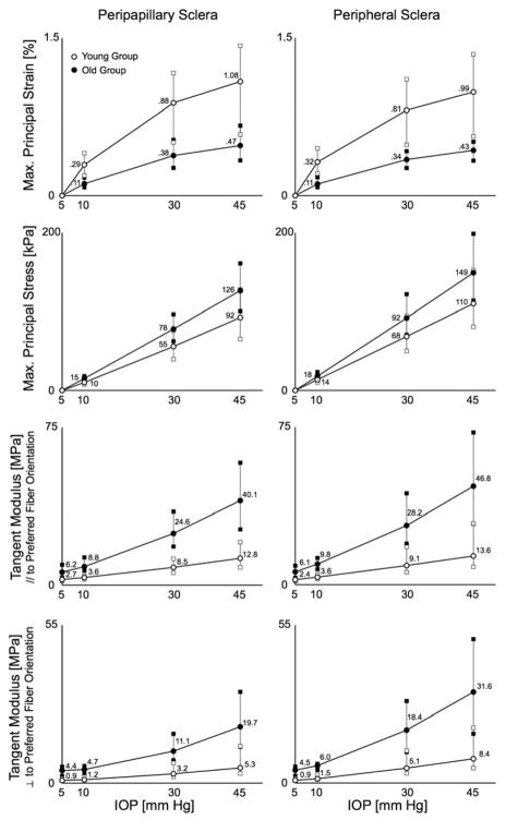
Purpose: To investigate the age-related differences in the inhomogeneous, anisotropic, nonlinear biomechanical properties of posterior sclera from old (22.9 +/- 5.3 years) and young (1.5 +/- 0.7 years) rhesus monkeys.

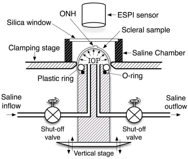
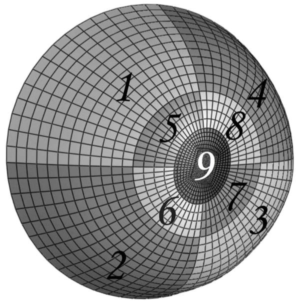
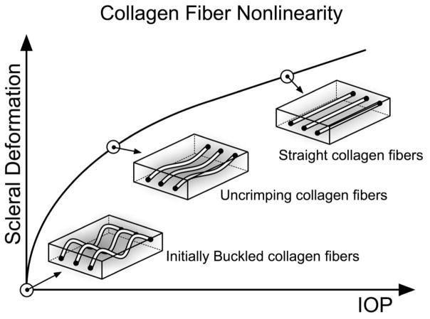
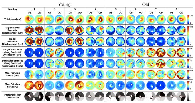
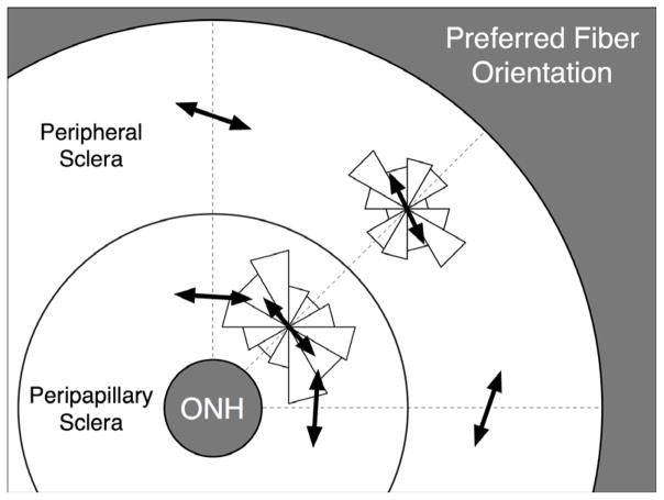
Methods: The posterior scleral shell of each eye was mounted on a custom-built pressurization apparatus, then intraocular pressure (IOP) was elevated from 5 to 45 mm Hg while the 3D displacements of the scleral surface were measured with speckle interferometry. Each scleral shell's geometry was digitally reconstructed from data generated by a 3-D digitizer (topography) and 20-MHz ultrasound (thickness). An inverse finite element (FE) method incorporating a fiber-reinforced constitutive model was used to extract a unique set of biomechanical properties for each eye. Displacements, thickness, stress, strain, tangent modulus, structural stiffness, and preferred collagen fiber orientation were mapped for each posterior sclera.

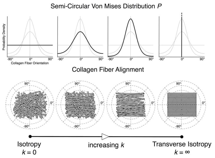
Results: The model yielded 3-D deformations of posterior sclera that matched well with those observed experimentally. The posterior sclera exhibited inhomogeneous, anisotropic, nonlinear mechanical behavior. The sclera was significantly thinner (P = 0.038) and tangent modulus and structural stiffness were significantly higher in old monkeys (P < 0.0001). On average, scleral collagen fibers were circumferentially oriented around the optic nerve head (ONH). No difference was found in the preferred collagen fiber orientation and fiber concentration factor between age groups.

Conclusions: Posterior sclera of old monkeys is significantly stiffer than that of young monkeys and is therefore subject to higher stresses but lower strains at all levels of IOP. Age-related stiffening of the sclera may significantly influence ONH biomechanics and potentially contribute to age-related susceptibility to glaucomatous vision loss.


Figures

Figure 1
Figure 1
Schematic of the scleral shell pressurization apparatus. The posterior scleral shell was first mounted onto the plastic ring, and then clamped slightly above the equator by moving the vertical stage toward the clamping stage. Saline outflow was interrupted after saline filled the posterior shell cavity and IOP reached 5 mmHg. The scleral surface was imaged with an electronic speckle pattern interferometry (ESPI) sensor as IOP increased from 5 to 45 mmHg in 0.2 mmHg increments.
Figure 2
Figure 2
Anatomically accurate geometry of one posterior scleral shell (from the clamping boundary to the ONH) that was reconstructed from experimental topography and thickness measurements. Regions 1–4 encompass the peripheral sclera, Regions 5–8 the peripapillary sclera and Region 9 the ONH. The peripapillary sclera (Regions 5–8) extended approximately 1.5 to 1.7 mm from the scleral canal, which was defined as the border between the ONH (Region 9) and the peripapillary sclera (Regions 5–8). The clamping ring was located approximately 3 mm posterior to the equator.
Figure 3
Figure 3
Semi-circular von-Mises distribution describing local collagen fiber alignment. As the fiber concentration factor k increases, collagen fibers become more aligned along the preferred fiber orientation (θp = 0° in this example). When k = 0, collagen fibers are randomly organized, resulting in equal stiffness in all orientations (relevant to skin tissue). This material symmetry is known as planar isotropy. When k = ∞, collagen fibers are all oriented in a particular preferred orientation, which creates high stiffness along θp and high compliance perpendicular to θp (relevant to tendons and ligaments). This material symmetry is known as transverse isotropy.
Figure 4
Figure 4
Uncrimping of the collagen fibers induces scleral stiffening at the macroscopic level. Initially the collagen fibers are buckled, then uncrimp and eventually become straight due to acute elevations of IOP, thus limiting scleral deformations at high IOP values. Note that the parameters c3 and c4 govern the degree of nonlinearity of each scleral shell.
Figure 5
Figure 5
Individual results for all posterior scleral shells as viewed from the back of the eye (Superior is up). Scleral thickness was experimentally measured at IOP = 5 mmHg and interpolated to obtain continuous thickness maps. Tangent modulus, structural stiffness, maximum principal stress and strain are shown for all eyes at a single IOP of 30 mmHg. Good agreement is observed between FE-computed and experimentally measured posterior displacements (plotted for an IOP range of 5–30 mmHg). Finally the preferred fiber orientation is shown for all eight regions of each eye, where // (black) corresponds to a collagen fiber organization tangent to the scleral canal (circumferential, θp = 0°) and ⊥ (white, θp = 90°) corresponds to a fiber organization that is perpendicular to the scleral canal (meridional). Note that the data for the two eyes of each monkey are much more similar than between monkeys, and there are clear age-related differences in all measures except for preferred collagen fiber orientation.
Figure 6
Figure 6
Pooled distributions (all eyes) of the preferred fiber orientation in both the peripapillary and peripheral scleral regions are shown as two symmetric rose diagrams, in which larger triangles indicate the most commonly derived orientations. Mean preferred fiber orientations (black arrows) were computed for each of both distributions using a circular statistics formula and were equal to 176.2° and 162.0° in the peripapillary and peripheral sclera, respectively. Note that 0° or 180° correspond to an orientation tangent to the scleral canal and 90° to an orientation perpendicular to the scleral canal. On average, this result suggests a tendency toward a circumferential organization of the collagen fibers around the scleral canal in both the peripapillary and the peripheral sclera.
Figure 7
Figure 7
Maximum principal strain, maximum principal stress, and tangent moduli distributions (error bars show the 25th, 50th, and 75th percentiles) plotted by age group for both scleral regions (peripapillary and peripheral) at the following IOPs: 5, 10, 30 and 45 mmHg. On average, sclera from the old monkeys exhibited higher tangent moduli and stress, but lower strain than that from the young monkeys. Note the nonlinear relationship between IOP and strain, which is due to the increase of tangent moduli with IOP (the sclera stiffens as IOP increases).

References

    1. Resnikoff S, Pascolini D, Etya’ale D, et al. Global data on visual impairment in the year 2002. Bull World Health Organ. 2004;82:844–851. - PMC - PubMed
    1. Foster A, Resnikoff S. The impact of Vision 2020 on global blindness. Eye. 2005;19:1133–1135. - PubMed
    1. Quigley H, Anderson DR. The dynamics and location of axonal transport blockade by acute intraocular pressure elevation in primate optic nerve. Invest Ophthalmol. 1976;15:606–616. - PubMed
    1. Quigley HA, Addicks EM. Chronic experimental glaucoma in primates. II. Effect of extended intraocular pressure elevation on optic nerve head and axonal transport. Invest Ophthalmol Vis Sci. 1980;19:137–152. - PubMed
    1. Bellezza AJ, Hart RT, Burgoyne CF. The optic nerve head as a biomechanical structure: initial finite element modeling. Invest Ophthalmol Vis Sci. 2000;41:2991–3000. - PubMed

Publication types