Case Reports
. 2009 Jun 4;360(23):2438-44.
doi: 10.1056/NEJMoa0809568.

An autoinflammatory disease due to homozygous deletion of the IL1RN locus

Affiliations
Case Reports

An autoinflammatory disease due to homozygous deletion of the IL1RN locus

Sreelatha Reddy et al. N Engl J Med. .

Abstract

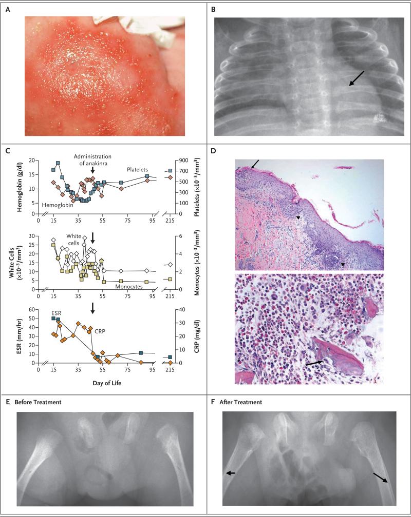
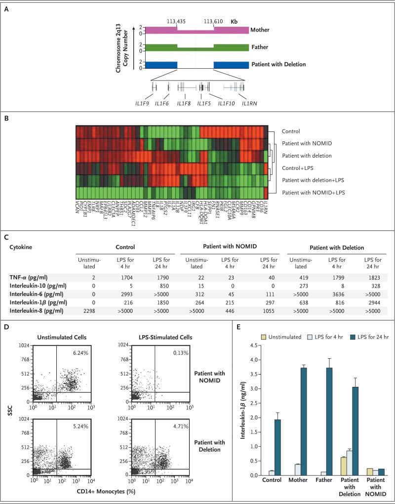
We describe a patient with an autoinflammatory disease in which the main clinical features are pustular rash, marked osteopenia, lytic bone lesions, respiratory insufficiency, and thrombosis. Genetic studies revealed a 175-kb homozygous deletion at chromosome 2q13, which encompasses several interleukin-1 family members, including the gene encoding the interleukin-1-receptor antagonist (IL1RN). Mononuclear cells, obtained from the patient and cultured, produced large amounts of inflammatory cytokines, with increasing amounts secreted after stimulation with lipopolysaccharide. A similar increase was not observed in peripheral-blood mononuclear cells from a patient with neonatal-onset multisystem inflammatory disorder (NOMID). Treatment with anakinra completely resolved the symptoms and lesions.


Figures

Figure 1
Figure 1. Clinical Phenotype of Our Patient with the Homozygous Deletion at the IL1RN Locus
The patient's rash consisted of erythematous macules with pustules (Panel A). Chest radiography showed osteopenia and anterior rib flaring with osteolytic lesions (Panel B, arrow). Panel C shows laboratory values before and after the administration of anakinra. In Panel D (hematoxylin and eosin), a skin-biopsy specimen (top) shows subcorneal pustules (arrow), filled with neutrophils, and focal acantholysis. Neutrophils also infiltrated and destroyed the infundibulum of hair follicles (arrowheads). A bone-biopsy specimen (bottom) shows fragments of woven bone with scalloping of the edges and a focally prominent cement line. Osteoclasts are scattered throughout, including one at a scalloped edge of the bone (arrow). Radiographs of the femurs before (Panel E) and after (Panel F) administration of anakinra show healing bone, as well as periosteal reactions (arrows) in response to treatment. CRP denotes C-reactive protein, and ESR erythrocyte sedimentation rate.
Figure 2
Figure 2. Genetic and Gene-Expression Characteristics of Our Patient with a Deletion at the IL1RN Locus and a Patient with Neonatal-Onset Multisystem Inflammatory Disease (NOMID)
Panel A shows the variation in genomic copy number in chromosome 2q13 for the patient and his mother and father, showing a homozygous 175-kb deletion in the patient and heterozygosity in his parents. The deletion includes genes encoding six members of the interleukin-1 family: the interleukin-1–receptor antagonist (IL1RN) and interleukin-1 family members 5, 6, 8, 9, and 10 (IL1F5, IL1F6, IL1F8, IL1F9, and IL1F10, respectively). The gene-expression profiles of peripheral-blood mononuclear cells from a control, our patient, and a patient with NOMID are shown in Panel B, before and after stimulation with lipopolysaccharide (LPS) for 4 hours. Red represents an increase in transcription, and green a decrease, with the intensity of the color indicating the degree of increase or decrease. The dendogram to the right of the microarray reflects the relatedness of the gene signatures between the samples. Panel C shows levels of inflammatory proteins in unstimulated and LPS-stimulated peripheral-blood mononuclear cells from a control, our patient, and a patient with NOMID. Panel D shows the numbers of CD14+ monocytes in peripheral-blood mononuclear cells from a patient with NOMID and in our patient before and after stimulation with LPS for 24 hours. The percentages of CD14+ monocytes remaining in the cultures are shown, according to side-scatter (SSC) analysis. Panel E shows mean levels of interleukin-1β in unstimulated and LPS-stimulated peripheral-blood mononuclear cells from our patient, his mother and father, a patient with NOMID, and a control subject. T bars indicate the standard deviation (not visible on some bars). TNF-α denotes tumor necrosis factor α.

Comment in

References

    1. Hull KM, Shoham N, Chae JJ, Aksentijevich I, Kastner DL. The expanding spectrum of systemic autoinflammatory disorders and their rheumatic manifestations. Curr Opin Rheumatol. 2003;15:61–9. - PubMed
    1. Prieur AM, Griscelli C. Arthropathy with rash, chronic meningitis, eye lesions, and mental retardation. J Pediatr. 1981;99:79–83. - PubMed
    1. Prieur AM, Griscelli C, Lampert F, et al. A chronic, infantile, neurological, cutaneous and articular (CINCA) syndrome: a specific entity analysed in 30 patients. Scand J Rheumatol Suppl. 1987;66:57–68. - PubMed
    1. Aksentijevich I, Nowak M, Mallah M, et al. De novo CIAS1 mutations, cytokine activation, and evidence for genetic heterogeneity in patients with neonatal-onset multi-system inflammatory disease (NOMID): a new member of the expanding family of pyrin-associated autoinflammatory diseases. Arthritis Rheum. 2002;46:3340–8. - PMC - PubMed
    1. Hoffman HM, Mueller JL, Broide DH, Wanderer AA, Kolodner RD. Mutation of a new gene encoding a putative pyrin-like protein causes familial cold autoinflammatory syndrome and Muckle-Wells syndrome. Nat Genet. 2001;29:301–5. - PMC - PubMed

Publication types

MeSH terms

Substances