. 2009 Jun 15;182(12):7353-63.
doi: 10.4049/jimmunol.0900657.

IL-10 deficiency unleashes an influenza-specific Th17 response and enhances survival against high-dose challenge

Affiliations

IL-10 deficiency unleashes an influenza-specific Th17 response and enhances survival against high-dose challenge

K Kai McKinstry et al. J Immunol. .

Abstract

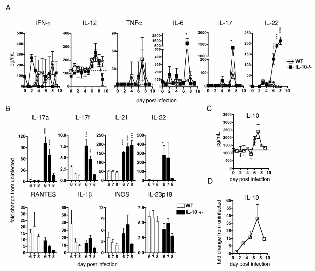
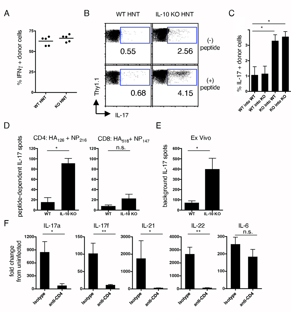
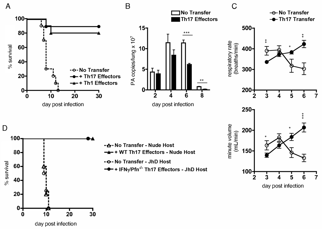
We examined the expression and influence of IL-10 during influenza infection. We found that IL-10 does not impact sublethal infection, heterosubtypic immunity, or the maintenance of long-lived influenza Ag depots. However, IL-10-deficient mice display dramatically increased survival compared with wild-type mice when challenged with lethal doses of virus, correlating with increased expression of several Th17-associated cytokines in the lungs of IL-10-deficient mice during the peak of infection, but not with unchecked inflammation or with increased cellular responses. Foxp3(-) CD4 T cell effectors at the site of infection represent the most abundant source of IL-10 in wild-type mice during high-dose influenza infection, and the majority of these cells coproduce IFN-gamma. Finally, compared with predominant Th1 responses in wild-type mice, virus-specific T cell responses in the absence of IL-10 display a strong Th17 component in addition to a strong Th1 response and we show that Th17-polarized CD4 T cell effectors can protect naive mice against an otherwise lethal influenza challenge and utilize unique mechanisms to do so. Our results show that IL-10 expression inhibits development of Th17 responses during influenza infection and that this is correlated with compromised protection during high-dose primary, but not secondary, challenge.


Figures

Figure 1
Figure 1
IL-10 expression negatively influences high-dose primary flu challenge. Age-matched WT or IL-10 KO mice were challenged with 1LD50 A/PR8 (5000 EID50). (A) weight loss and conditional survival (n=15 mice / group, and * = P < 0.05 from 1 of 5 similar experiments (exp)). (B) WT mice infected with 1 LD50 A/PR8 were treated with either IL-10 receptor blocking antibody or an isotype control, as described, and weight loss and conditional survival monitored (n=8, and * = P < 0.05 from 1 of 2 independent exp). Weight loss in A-B is of all surviving animals. (C) On stated days post-infection, viral titers were determined by quantitative PCR (n=5 / group / day from 1 of 3 similar exp), and (D) respiratory rates and minute volumes monitored (n= at least 7, * = P < 0.05, and *** = P < 0.001 from 1 of 2 independent exp). (E) H&E stained lung sections were scored blindly for levels of immunopathology (n=3–7 mice / group / day from 1 of 2 exp).
Figure 2
Figure 2
Increased expression of Th17-associated cytokines in the absence of IL-10. WT or IL-10 KO mice were challenged with 1LD50 A/PR8 and on stated days, lungs isolated and analyzed for (A and C) protein, or (B and D) message for stated cytokines, as described (n=3 mice / group / time point for A and C; n=5 for B and D). Dotted lines in A and C represent average level of protein in uninfected mice. Data presented is representative of two similar independent exp separately measuring protein and message (* = P < 0.05, ** = P < 0.01, and *** = P < 0.001).
Figure 3
Figure 3
Effector CD4 T cells co-producing IFNγ are the primary source of IL-10 during flu infection. (A) WT mice were treated with depleting or isotype control antibodies and infected with 1 LD50 A/PR8. On d7, lungs were analyzed for IL-10 expression (n=5, and * = P < 0.05). (B) 2×10⁶ naïve HNT.Thy1.1 CD4 T cells were transferred to WT hosts then infected with 1 LD50 A/PR8, and IL-10+ donor cells determined by ICCS on d7 (n=5). (C) Representative examples of donor cell IL-10/IFNγ and isotype staining +/− peptide stimulation. (D) Stated numbers of donor cells were transferred and ICCS performed as in (C); the ratio of IFNγ+/IL-10 to IFNγ+/IL-10+ donor cells in lungs is shown (n=3). (E) Representative example of IL-10/IFNγ staining gated on CD4 T cells from DLN and lung of WT mice in the absence of adoptive transfer. (F) CD25/FoxP3 staining of host and donor lung CD4 T cells on d 7 post-infection. (G) Total IL-10+ T cells from FoxP3.GFP mice were separated on the basis of GFP expression (* = P < 0.05). Data is representative of 2 independent exp for A, D, G and greater than 5 for B, C, E, F.
Figure 4
Figure 4
Th1/Th17 phenotype of responding flu-specific IL-10 KO T cells. 2×10⁶ naïve WT or IL-10 KO HNT cells were transferred to WT or IL-10 KO hosts, respectively, then infected with 1LD50 A/PR8. 7 d post-challenge, lung HNT cells were analyzed for (A) IFNγ or (B) IL-17 +/− peptide stimulation (n=5). (C) Peptide-dependent IL-17 production from WT or IL-10 KO HNT cells transferred to WT or IL-10 KO hosts (n=3, and * = P < 0.05). WT or IL-10 KO mice were challenged with 1LD50 A/PR8 and ELISPOT analysis performed on d 7 for IL-17. (D) CD4 and CD8 peptide-specific, and (E) non-antigen elicited IL-17 spots per lung (n=5, and * = P < 0.05). (F) WT mice were treated with CD4 depleting or isotype control antibodies and infected with 1 LD50 A/PR8. On d 7, lungs were analyzed for stated cytokines (n=5, * = P < 0.05 and ** = P < 0.01). A-C are representative of 3 independent exp, E-F of 2.
Figure 5
Figure 5
Th17-polarized effectors retain defining aspects of their phenotype in vivo following flu challenge. (A) IFNγ and IL-17 staining of Th1 and Th17 effectors +/− peptide stimulation, and (B) IFNγ, IL-4, IL-17, IL-21 and IL-22 measured in 24 h supernatants of indicated effectors stimulated with APC and peptide. (C) Message for T-bet, GATA-3, and ROR-γt in polarized effectors. (D) Granzyme B staining of Th1 and Th17 effectors prior to and d3 following adoptive transfer and flu infection. (E) IFNγ and IL-17 staining of Th1 and Th17 HNT effectors in lungs 3d after transfer and flu infection. A-B are representative of 4 independent exp, D-E of 3 similar exp.
Figure 6
Figure 6
Th17-polarized effectors protect against lethal flu challenge employing novel mechanisms. 5×10⁶ Th1 or Th17 HNT effectors were transferred to naïve BALB/c mice subsequently infected with 2 LD50 A/PR8 (10,000 EID50) and (A) conditional survival monitored (n=10 mice per group). (B) On stated days, viral titers were determined by quantitative PCR (n=5 mice / group / day, ** = P < 0.01, and *** = P < 0.001). (C) Respiratory rate and minute volume was determined as described (n=10 mice / group / time-point, * = P < 0.05, ** = P < 0.01, and *** = P < 0.001). (D) 5×10⁶ Th17 effectors generated from WT or INFγ-/Perforin KO HNT cells were transferred to nude or JHD hosts, respectively. All mice were subsequently infected with a lethal dose (1500 EID50) of A/PR8. Conditional survival is shown (n=10 mice / group). A-C representative of at least 2 independent exp, D is a summary of 2 independent exp for each condition, with n=5 mice per group.
Figure 7
Figure 7
Reduced IL-10 expression during secondary flu responses. (A) WT or IL-10 KO mice were infected with 0.1LD50 A/PR8 (H1N1) and on stated days post primary infection, mice were challenged with 300 LD50 A/Philippines (H3N2) and monitored for weight loss (n=5 mice / group). No primed mice succumbed to secondary challenge. (B) After naïve HNT cell transfer, lung and dLN resident donor cells were assayed for IL-10 production by ICCS on stated days post 0.1LD50 A/PR8 challenge. (C) 2×10⁶ naïve or memory HNT cells (Th1-polarized rested effectors) were transferred to naïve hosts subsequently infected with 1 LD50 A/PR8. On stated days, lungs (n=3 / group) were analyzed for IL-10 message. (D) Lung homogenates (n=3 mice / day) from naïve mice infected with 1 LD50 A/PR8 and from mice primed 60 days previously with A/PR8 and challenged with 300 LD50 A/Philippines were analyzed for IL-10 expression. Data in B-D is representative of 2 independent exp.

References

    1. Fiorentino DF, Bond MW, Mosmann TR. Two types of mouse T helper cell. IV. Th2 clones secrete a factor that inhibits cytokine production by Th1 clones. J Exp Med. 1989;170:2081–2095. - PMC - PubMed
    1. Moore KW, de Waal Malefyt R, Coffman RL, O'Garra A. Interleukin-10 and the interleukin-10 receptor. Annu Rev Immunol. 2001;19:683–765. - PubMed
    1. Couper KN, Blount DG, Riley EM. IL-10: the master regulator of immunity to infection. J Immunol. 2008;180:5771–5777. - PubMed
    1. Li C, Corraliza I, Langhorne J. A defect in interleukin-10 leads to enhanced malarial disease in Plasmodium chabaudi chabaudi infection in mice. Infect Immun. 1999;67:4435–4442. - PMC - PubMed
    1. Hunter CA, Ellis-Neyes LA, Slifer T, Kanaly S, Grunig G, Fort M, Rennick D, Araujo FG. IL-10 is required to prevent immune hyperactivity during infection with Trypanosoma cruzi. J Immunol. 1997;158:3311–3316. - PubMed

Publication types

MeSH terms