. 2009 Jun 15;182(12):7415-21.
doi: 10.4049/jimmunol.0804214.

Modulation of p38 MAPK activity in regulatory T cells after tolerance with anti-DNA Ig peptide in (NZB x NZW)F1 lupus mice

Affiliations

Modulation of p38 MAPK activity in regulatory T cells after tolerance with anti-DNA Ig peptide in (NZB x NZW)F1 lupus mice

Elaine V Lourenço et al. J Immunol. .

Abstract

Treatment of (NZB x NZW)F(1) (NZB/W) lupus-prone mice with the anti-DNA Ig-based peptide pConsensus prolongs the survival of treated animals and effectively delays the appearance of autoantibodies and glomerulonephritis. We have previously shown that part of these protective effects associated with the induction of CD4(+)CD25(+)Foxp3(+) regulatory T cells (Tregs) that suppressed autoantibody responses. Because the effects of pConsensus appeared secondary to qualitative rather than quantitative changes in Tregs, we investigated the molecular events induced by tolerance in Tregs and found that signaling pathways including ZAP70, p27, STAT1, STAT3, STAT6, SAPK, ERK, and JNK were not significantly affected. However, peptide tolerization affected in Tregs the activity of the MAPK p38, whose phosphorylation was reduced by tolerance. The pharmacologic inhibition of p38 with the pyridinyl imidazole inhibitor SB203580 in naive NZB/W mice reproduced in vivo the effects of peptide-induced tolerance and protected mice from lupus-like disease. Transfer experiments confirmed the role of p38 in Tregs on disease activity in the NZB/W mice. These data indicate that the modulation of p38 activity in lupus Tregs can significantly influence the disease activity.


Figures

Figure 1
Figure 1. Effects of peptide tolerization on in vivo numbers of Tregs
NZB/W mice tolerized with pCons have similar percentage (a) and total (b) numbers of Tregs than age- and weight-matched controls (8 to 12 mice per group). P is not significant between tolerized mice and controls for both panels a and b.
Figure 2
Figure 2. Tregs from tolerized mice confer higher protection than control Tregs after transfer into syngeneic mice
The transfer of equal numbers of purified NZB/W Tregs from pCons-tolerized or control mice into syngeneic 16 weeks-old recipient mice associates with a delayed development of renal disease (as manifested by proteinuria, top; *P<0.04), and an increased survival (bottom; *P<0.001) of recipients of pCons-derived Tregs than recipients of control Tregs.
Figure 3
Figure 3. Signalling events after tolerization
Immunoblots for phosphorylated (p-) and non-phosphorylated ZAP-70, p27, ERK, JNK, SAPK, STAT1, STAT3, STAT6, and p38 in sorted Tregs from control mice (ctrl) and from mice tolerized (tol) with pCons peptide show a reduced phosphorylation of p38 in Tregs from tolerized mice. In the quadrant, a direct comparison of phosphorylated p38 to non-phosphorylated p38 between purified CD4+CD25+ T cells and CD4+CD25 T cells shows that modulated activation of p38 occurs in Tregs after tolerance but not in effector T cells, the latter displaying virtually absent basal activation of p38. Graphs show the densitometric quantitation of protein to the housekeeping gene (p27) and to the non-phosphorylated form (all others). The results were reconfirmed in four to seven independent experiments and using different Ab from two different commercial suppliers (see Materials and Methods). There was no difference in Foxp3 expression by immunoblot in sorted Tregs from control and tolerized mice (not shown).
Figure 4
Figure 4. Effects of tolerization with peptide on p38 activity in NZB/W Tregs
a. Flow cytometry analysis for the intracellular expression of p38 (left panel) and p-p38 (right panel) in gated Tregs (CD4+CD25+Foxp3+cells) from tolerized (gray line) and control mice (black line). b. Tregs from pCons-tolerized mice have similar mean fluorescence intensity of non-phosphorylated p38 (P ns) and reduced phosphorylation of p38 than controls (n = 8 per group; P<0.004). c. Cumulative data on the intracellular expression of p38 and p-p38 detected by flow cytometry in CD4+CD25+Foxp3+ T cells from tolerized (n = 8) and control (n = 8) mice; P<0.01.
Figure 5
Figure 5. Inhibition of p38 delays lupus-like disease in NZB/W mice
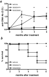
Sixteen- to twenty-week old premorbid mice (n = 6 per group) were treated with p38 inhibitor SB203580, control SB202474, or vehicle as indicated in the Materials and Methods and then monitored for serum anti-DNA (a; *P<0.04) and survival (b; *P<0.003).
Figure 6
Figure 6. In vivo inhibition of p38 in NZB/W mice associates with a reduced serum concentration of inflammatory cytokines
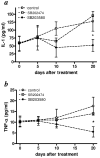
Age matched (12 to 16 weeks-old) NZB/W mice (n = 6 per group) were treated with the p38 inhibitor SB203580, SB202474 control, or vehicle, as indicated in the Materials and Methods. Serum concentration of IL-1 (a) and TNF-α (b) were monitored by ELISA; *P<0.05.
Figure 7
Figure 7. Effects of SB203580 and pCons on p38 inhibition in Tregs
a. Ratio of phosphorylated p-p38 to total p38 detected by intracellular flow cytometry in sorted CD4+CD25+Foxp3+ T cells from tolerized (n = 5-8) and control mice (n = 5-8) treated with 2 mg/kg/day i.p. for 2 weeks with SB203580, SB202474, or vehicle (*P<0.04; **P<0.01) (b) Numbers of Tregs after treatment (P not significant).
Figure 8
Figure 8. Effects of in vivo p38 inhibition in Tregs
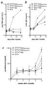
Irradiated NZB/W mice received 2 × 10⁶ purified Tregs from syngeneic mice tolerized with pCons that had been treated for 2 weeks i.p. with 2 mg/kg/day SB203580 (TregsSB203580), SB202474 (TregsSB202474), or vehicle, together with 2 × 10⁶ purified CD4+CD25 T helper cells (Th) and 2 × 10⁶ purified B cells (1:1:1 ratio Tregs:Th:B). Control mice received equal numbers of the individual cell subsets (not shown) or a combination of Th and B cells. Recipient mice (n = 6 per group) were monitored for serum anti-DNA IgG (a), total IgG (b) and development of proteinuria (c). TregsSB203580/Th/B vs. controls, *P <0.05.

References

    1. Sakaguchi S, Sakaguchi N, Asano M, Itoh M, Toda M. Immunologic self-tolerance maintained by activated T cells expressing IL-2 receptor α-chains (CD25). Breakdown of a single mechanism of self-tolerance causes various autoimmune diseases. J. Immunol. 1995;155:1151–1164. - PubMed
    1. Asano M, Toda M, Sakaguchi N, Sakaguchi S. Autoimmune disease as a consequence of developmental abnormality of a T cell subpopulation. J. Exp. Med. 1996;184:387–396. - PMC - PubMed
    1. Sakaguchi S. Naturally arising CD4+ regulatory T cells for immunologic self-tolerance and negative control of immune responses. Annu. Rev. Immunol. 2004;22:531–562. - PubMed
    1. Shevach EM. CD4+CD25+ suppressor T cells: more questions than answers. Nat. Rev. Immunol. 2002;2:389–400. - PubMed
    1. La Cava A. T-regulatory cells in systemic lupus erythematosus. Lupus. 2008;17:421–425. - PubMed

Publication types

MeSH terms