. 2009 Jun 15;182(12):7422-9.
doi: 10.4049/jimmunol.0900075.

The immunomodulatory action of sialostatin L on dendritic cells reveals its potential to interfere with autoimmunity

Affiliations

The immunomodulatory action of sialostatin L on dendritic cells reveals its potential to interfere with autoimmunity

Anderson Sá-Nunes et al. J Immunol. .

Abstract

Sialostatin L (SialoL) is a secreted cysteine protease inhibitor identified in the salivary glands of the Lyme disease vector Ixodes scapularis. In this study, we reveal the mechanisms of SialoL immunomodulatory actions on the vertebrate host. LPS-induced maturation of dendritic cells from C57BL/6 mice was significantly reduced in the presence of SialoL. Although OVA degradation was not affected by the presence of SialoL in dendritic cell cultures, cathepsin S activity was partially inhibited, leading to an accumulation of a 10-kDa invariant chain intermediate in these cells. As a consequence, in vitro Ag-specific CD4(+) T cell proliferation was inhibited in a time-dependent manner by SialoL, and further studies engaging cathepsin S(-/-) or cathepsin L(-/-) dendritic cells confirmed that the immunomodulatory actions of SialoL are mediated by inhibition of cathepsin S. Moreover, mice treated with SialoL displayed decreased early T cell expansion and recall response upon antigenic stimulation. Finally, SialoL administration during the immunization phase of experimental autoimmune encephalomyelitis in mice significantly prevented disease symptoms, which was associated with impaired IFN-gamma and IL-17 production and specific T cell proliferation. These results illuminate the dual mechanism by which a human disease vector protein modulates vertebrate host immunity and reveals its potential in prevention of an autoimmune disease.


Figures
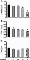
Figure 1
Figure 1
Concentration-response effect of SialoL on cytokine production by LPS-stimulated DCs. Enriched CD11c+ cells (105/well) were preincubated with increasing concentrations of SialoL for 2 h and subsequently stimulated with LPS (50 ng/ml). Supernatants were collected for IL-12p70 (A), TNF-α (B) and IL-10 (C) determination by ELISA. *P < 0.05 versus LPS group without SialoL.
Figure 2
Figure 2
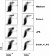
Sialo L suppresses LPS-induced CD80 and CD86 expression by DCs. Four million bone marrow-derived DCs were preincubated with medium or SialoL 3 μM for 2 h prior to an overnight incubation in the presence of LPS (50 ng/ml). Cells were stained with fluorochrome-labeled Abs specific for CD11c, CD80, CD86, and MHC class II, and analyzed by flow cytometry. The data shown were gated from live CD11c+ cells.
Figure 3
Figure 3
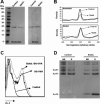
SialoL binds cathepsin S inside DCs leading to an accumulation of Ii-p10 intermediates. Ten million DCs were incubated with medium or SialoL 3 μM for 24 h. Cells were lysed and separated on a 12% SDS-PAGE gel, and transferred to nitrocellulose membranes. Cathepsin S was detected with specific antibodies in nonboiled and boiled samples (A). Densitometry of the bands from this representative experiment is displayed (B). SialoL does not inhibit OVA cleavage into DCs. Cells were preincubated for 3 h at 37°C in the presence or absence of 3 μM SialoL and further incubated with DQ™ OVA (1 μg/ml) for 2 h at 37°C. OVA fragmentation was analyzed by flow cytometry (C). DCs lysates were prepared as described above and Ii intermediates were detected with specific antibodies in nonboiled and boiled samples (D).
Figure 4
Figure 4
SialoL suppresses antigen-specific CD4+ T cell proliferation, but not mixed lymphocyte reaction. For antigen-specific proliferation, enriched CD11c+ cells (2.5 × 10⁴/well) from 6-d BM cultures were preincubated with SialoL 3 μM for 0, 1 or 3 h, pulsed with OVA (1 μg/ml) and cultured with purified CD4+ T cells (105/well) for 72 h (A). For mixed lymphocyte reaction, total spleen cells (2.5 × 10⁵/well) from BALB/c mice were preincubated with SialoL in different concentrations for 3 h, and total spleen cells from OT-II mice (2.5 × 10⁵/well) were added and cultured for 72 h (B). Proliferation was measured as described in Material and Methods. *P < 0.05 versus OVA group without SialoL preincubation.
Figure 5
Figure 5
Cathepsin S accounts for the inhibition of T cell proliferation in SialoL pretreated cultures. Enriched CD11c+ cells (2.5 × 10⁴/well) from 6-d BM cultures of cathepsin L−/− and S−/− mice and their respective WT littermates were preincubated with medium or SialoL 3 μM for 3 h, pulsed with OVA (1 μg/ml), and cultured with purified CD4+ T cells (105/well) for 72 h. WT and cathepsin L−/− cultures (A). WT and cathepsin S−/− cultures (B). Proliferation was measured as described. *P < 0.05 versus respective medium groups; #P < 0.05 versus respective WT groups.
Figure 6
Figure 6
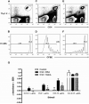
SialoL treatment impairs in vivo proliferation and recall response. Adoptive transfer experiment. B6.PL-Thy1a/CyJ mice were i.v. injected with 2.5 × 10⁶ CD4+ T cells from OT-II mice labeled with CFSE. One day later, mice were immunized in two sites of the flanks with 10 μg OVA emulsified in CFA and treated with BSA or SialoL as described. After 96 h, the LNs were removed and proliferation was measured by flow cytometry based on CFSE dilution of gated CD4+/Thy1.2+ cells. Non-immunized mice (A, B), BSA-treated mice immunized with OVA (C, D), SialoL-treated mice immunized with OVA (E, F). Recall response. C57BL/6 naïve mice were immunized in two sites of the flanks with 10 μg OVA emulsified in CFA and treated with BSA or SialoL as described. Thirty days later, LN cells were stimulated in vitro with increasing concentrations of OVA or Con A for 72 h and proliferation was measured (G). *P < 0.05 versus OVA + BSA group.
Figure 7
Figure 7
Resistance of SialoL treated mice to EAE is associated with impaired proliferation and cytokine production upon in vitro restimulation with MOG. Time course of the clinical score from C57BL/6 mice immunized with MOG (35−55) and treated with BSA or SialoL as described (n= 5−11 mice/group) (A). Ten days after immunization, LN cells from both groups were restimulated in vitro with 1 and 5 μg/ml of MOG for 72 h. Proliferation was measured (B) and levels of IFN-γ (C) and IL-17 (D) were determined in the culture supernatants by ELISA. *P < 0.05 versus PBS group; #P < 0.05 versus SialoL2 group; &P < 0.05 versus MOG/BSA group.

References

    1. Titus RG, Ribeiro JM. The role of vector saliva in transmission of arthropod-borne disease. Parasitol Today. 1990;6:157–160. - PubMed
    1. Hill CA, Wikel SK. The Ixodes scapularis Genome Project: an opportunity for advancing tick research. Trends Parasitol. 2005;21:151–153. - PubMed
    1. Center for Disease Control and Prevention. Lyme disease - United States, 2001−2002. Morb Mortal Wkly Rep. 2004;53:107–113.
    1. Ramachandra RN, Wikel SK. Modulation of host-immune responses by ticks (Acari: Ixodidae): effect of salivary gland extracts on host macrophages and lymphocyte cytokine production. J Med Entomol. 1992;29:818–826. - PubMed
    1. Zeidner N, Mbow ML, Dolan M, Massung R, Baca E, Piesman J. Effects of Ixodes scapularis and Borrelia burgdorferi on modulation of the host immune response: induction of a TH2 cytokine response in Lyme disease-susceptible (C3H/HeJ) mice but not in disease-resistant (BALB/c) mice. Infect Immun. 1997;65:3100–3106. - PMC - PubMed

Publication types

MeSH terms