. 2009 Jun 15;182(12):7708-17.
doi: 10.4049/jimmunol.0900666.

Analogous interactions in initiating complexes of the classical and lectin pathways of complement

Affiliations

Analogous interactions in initiating complexes of the classical and lectin pathways of complement

Anna E Phillips et al. J Immunol. .

Abstract

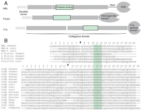
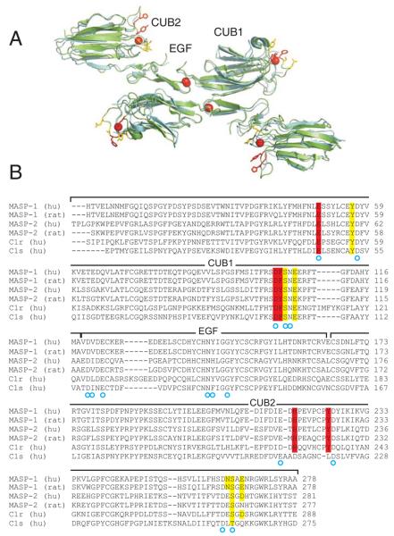
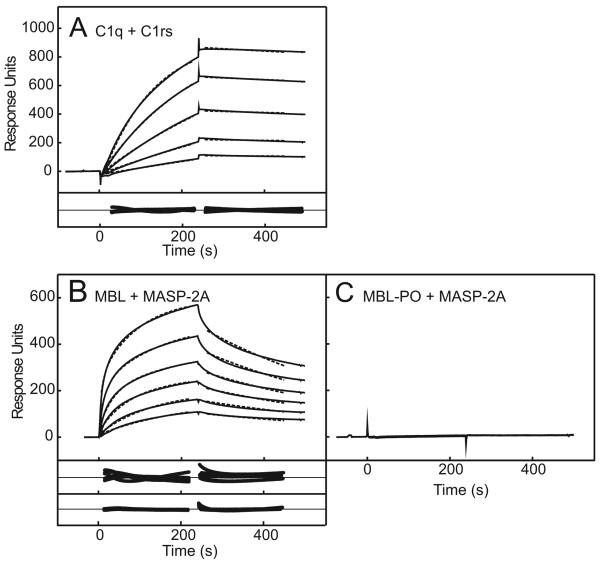
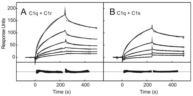
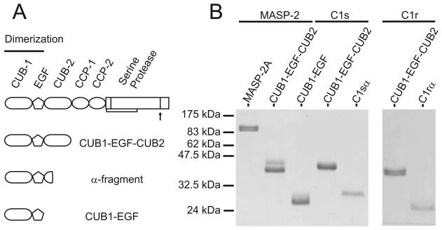
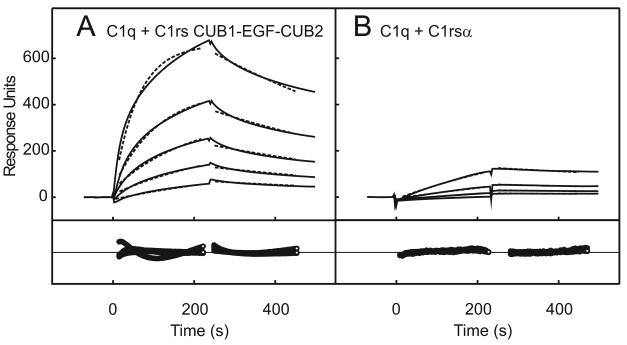
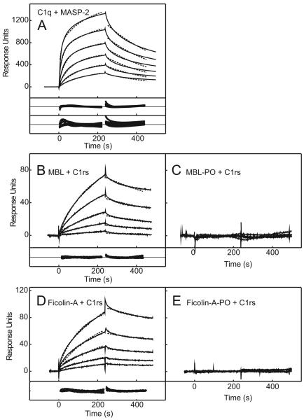
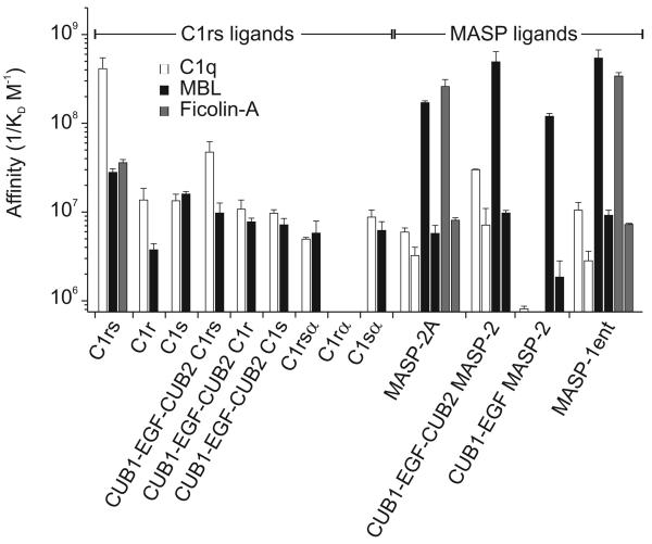
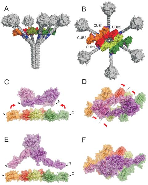
The classical and lectin pathways of complement activation neutralize pathogens and stimulate key immunological processes. Both pathways are initiated by collagen-containing, soluble pattern recognition molecules associated with specific serine proteases. In the classical pathway, C1q binds to Ab-Ag complexes or bacterial surfaces to activate C1r and C1s. In the lectin pathway, mannan-binding lectin and ficolins bind to carbohydrates on pathogens to activate mannan-binding lectin-associated serine protease 2. To characterize the interactions leading to classical pathway activation, we have analyzed binding between human C1q, C1r, and C1s, which associate to form C1, using full-length and truncated protease components. We show that C1r and C1s bind to C1q independently. The CUB1-epidermal growth factor fragments contribute most toward binding, but CUB2 of C1r, but not of C1s, is also important. Each C1rs tetramer presents a total of six binding sites, one for each of the collagenous domains of C1q. We also demonstrate that subcomponents of the lectin and classical pathways cross-interact. Thus, although the stoichiometries of complexes differ, interactions are analogous, with equivalent contacts between recognition and protease subcomponents. Importantly, these new data are contrary to existing models of C1 and enable us to propose a new model using mannan-binding lectin-mannan-binding lectin-associated serine protease interactions as a template.


Figures

FIGURE 1
FIGURE 1
Aligned sequences of the collagenous domains of MBLs, ficolins and C1qs. A, Domain organisations of MBL, ficolins and C1q. The carbohydrate-recognition domain of MBL is labelled as CRD. The putative protease-binding site (MASP or C1r/C1s) is indicated. B, Aligned sequences of the collagenous domains of MBLs, ficolins and C1qs. The protease-binding motif is shaded. Numbering of amino acid triplets is based upon the sequences of human MBL and C1q. The positions of the kinks in the collagenous domains of MBLs, rat ficolin-A and C1qs are shown by arrows. Positions of hydroxyproline (O) and hydroxylysine residues (underlined) are based upon the sequences of rat MBL (12) and human C1q (50).
FIGURE 2
FIGURE 2
Structures of the CUB1-EGF-CUB2 fragments of human MASP-1 (blue) and rat MASP-2 (green). A, Residues implicated in binding to MBL in MASP-1 are highlighted in red (highest contributions to binding) and yellow (lesser, but significant contributions to binding) (10). Ca2+ are shown as red spheres. In the rat structure, the Ca2+ binding site in each of the CUB domains is unoccupied and the loops forming the MBL-binding site are partially disordered, probably due to the conditions of crystallisation. The pdb codes for the MASP-1/-3 and MASP-2 structures are 3DEM (10) and 1NT0 (13), respectively. B, Aligned sequences of the CUB1-EGF-CUB-2 domains of MASPs, C1r and C1s. Residues implicated in MBL binding are highlighted in red and yellow (using the same colour scheme as in A, above). Coordinating ligands for the three Ca2+ (one in each CUB domain and one in the EGF-like domain) are indicated by blue circles.
FIGURE 3
FIGURE 3
Interactions between C1q and C1rs tetramers and between MBL and MASP-2 measured by surface plasmon resonance. A, C1q (~16,500 response units) was immobilized on to the surface of a sensor chip and C1rs was injected at 99, 49.3, 24.6, 12.3, 6.2 nM. Dotted lines show the fit to a single component binding model and the residuals to the fit are shown in the panel below. The amplitude of the residual plot is ± 120 response units. MASP-2A at 386, 193, 97, 48, 24 and 12 nM was injected over immobilised MBL in B, and MBL-PO in C; each at ~13,500 response units. The data for MBL were fitted to a single complex binding model and a two-complex parallel reaction binding model and the residuals are shown in the panels below the binding data. Amplitudes of the residual plots are ± 120 response units. The fit to the two-complex model is shown by the dotted line. The complex kinetics probably reflect binding of two MASPs to MBL trimers and tetramers, which has been detected previously at high concentrations of MASP-2 (14). In theory oligomeric heterogeneity of MBL, resulting in multiple MBL-MASP complexes forming on the chip surface, could also account for the observed kinetics. However, different MBL oligomers bind to MASP-2 with similar affinities (14), so this possibility can be discounted.
FIGURE 4
FIGURE 4
Binding of C1r and C1s to immobilised C1q by surface plasmon resonance. In A, C1r, at 500, 333, 167, 83, 42 and 21 nM and in B, C1s, at 333, 167, 83, 42 and 21 nM) were injected over immobilized C1q (~16,500 response units). Data were fitted to single complex binding models and the fits are shown by the dotted lines. Amplitudes of the residual plots are ± 50 response units.
FIGURE 5
FIGURE 5
SDS-PAGE of full-length and truncated recombinant MASP-2 proteins, recombinant C1r and C1s fragments and purified α-fragments of C1r and C1s. A, Schematic representation of the domain organisations of C1r, C1s, MASP-2 and their N-terminal fragments. B, SDS-PAGE analysis of purified recombinant MASP proteins and fragments of C1r and C1s, separated on 15% gels and stained with Coomassie blue.
FIGURE 6
FIGURE 6
Binding of truncated C1rs complexes to C1q by surface plasmon resonance. A, Complexes comprising the CUB1-EGF-CUB2 fragments of C1rs (at 244, 122, 61, 31 and 15 nM) and B, α-fragments of C1rs (at 384, 192, 96 and 48 nM) were injected over immobilized C1q (~16,500 response units). Data were fitted to single complex binding models and the fits are shown by the dotted lines. The amplitudes of the residual plots are ± 120 and ± 20 response units in A and B, respectively.
FIGURE 7
FIGURE 7
Cross binding between C1q and MASP-2A, MBL and C1rs tetramers and Ficolin-A and C1rs tetramers measured by surface plasmon resonance. A, C1q (~18,500 response units) was immobilized on to the surface of a sensor chip and MASP-2A was injected at 440, 220, 110, 55, 28 nM. Dotted lines show the fit to a two-complex parallel-reaction binding model. The amplitudes of the residual plots are ± 300 response units. The binding kinetics probably reflects binding of two MASPs to each C1q molecule. This arrangement would be compatible with the known stoichiometry of the C1 complex, in which four protease protomers (i.e. a C1rs tetramer or two MASP dimers) are accommodated by C1q. B and C, C1rs tetramers were injected over MBL and MBL-PO (~13,500 response units each) at 99, 49.3, 24.6, 12.3, 6.2 nM. D and E, C1rs tetramers were injected over Ficolin-A and Ficolin-A-PO (~12,000 and 10,000 response units) at 266, 133, 67, 34 and 17 nM. The data in B and D were fitted to single complex binding models and the residuals (± 15 response units) are shown in the panels below the main graphs. Fits are shown by the dotted lines. In each case, the data fitted reasonably well to a single-site model, Nevertheless, the overall amplitude of each interaction was significantly lower than for binding to MASP-2A under similar conditions (see Fig. 3B for comparison), implying that only a subset of MBL or ficolin molecules were binding to C1rs tetramers (probably tetramers and trimers of subunits).
FIGURE 8
FIGURE 8
Affinities of the cognate and non-cognate interactions between subcomponents of the lectin and classical pathway initiating complexes. Binding affinities are expressed as 1/KD (M−1).
FIGURE 9
FIGURE 9
New model of the interactions between C1q and C1rs in the C1 complex. A, Side and B, top views of C1q and the CUB1-EGF-CUB2 domains of C1r and C1s. The homology model of the dimeric CUB1-EGF-CUB2 portions of C1r (orange and dark green) and C1s (red and pale green) was generated using the crystal structure of the corresponding domains of MASP-1/-3 (10), as described in Materials and methods. Two antiparallel C1rs heterodimers (red/orange and pale green/dark green) lie alongside each other in a planar arrangement, held together by the interactions with C1q subunits. The six binding sites for C1q (purple), one on each of the CUB domains of C1r and one on each CUB1 of C1s, simultaneously interact with the protease-binding motif on separate C1q subunits (blue). Side C and top D views of the C1rs CUB1-EGF-CUB2 portion aligned with the CCP1-CCP2-SP domains (purple) of zymogen C1r. C1q has been omitted for clarity. The distance between the C-termini of the C1r CUB2 domains is much larger (~180 Å) than that between the N-termini of the C1r CCP1 domains (~100 Å), suggesting that the CUB1-EBF-CUB2 portion of the C1rs tetramer must be distorted in the zymogen complex. The CCP1-CCP2-SP domains of C1s are not shown. Red arrows indicate the likely movements of the C1r CUB2 domains (in C) and the relative movement of each C1rs CUB1-EGF-CUB2 fragment (in D) that would permit connection to the CCP-CCP-SP domains in the zymogen complex. These changes in turn would pull the C1q subunits together, reducing the angle between subunits. The pdb code for the structure of the C1r CCP-CCP-SP zymogen complex is 1GPZ. Side (E) and top (F) views of the C1rs CUB1-EGF-CUB2 portions aligned with the CCP-CCP-SP domains of active C1r. Upon target recognition of the C1 complex by the globular heads of C1q, the collagenous subunits probably splay further apart again. This movement separates the C1r polypeptides and pulls them away from each other, so that the SP domain of one C1r chain can cleave the activation site on the other chain. One possible such arrangement is observed in symmetry-related chains within the crystal structure of the cleaved C1r CCP-CCP-SP fragments, in which the S1 site of one SP domain interacts with the P1 site of the other (pdb code: 1QY0; Kardos et al. (2008). In this structure, the distance between the N-termini of the C1r CCP1 domains has increased to ~165 Å, comparable to the distance between the C-termini of the C1r CUB2 domains.

Similar articles

Cited by

References

    1. Porter RR, Reid KBM. The biochemistry of complement. Nature. 1978;275:699–704. - PubMed
    1. Carroll MC. The complement system in regulation of adaptive immunity. Nat Immunol. 2004;5:981–986. - PubMed
    1. Gaboriaud C, Teillet F, Gregory LA, Thielens NM, Arlaud GJ. Assembly of C1 and the MBL- and ficolin-MASP complexes: structural insights. Immunobiology. 2007;212:279–288. - PubMed
    1. Girija UV, Dodds AW, Roscher S, Reid KB, Wallis R. Localization and Characterization of the Mannose-Binding Lectin (MBL)-Associated-Serine Protease-2 Binding Site in Rat Ficolin-A: Equivalent Binding Sites within the Collagenous Domains of MBLs and Ficolins. J Immunol. 2007;179:455–462. - PMC - PubMed
    1. Reid KB, Sim RB, Faiers AP. Inhibition of the reconstitution of the haemolytic activity of the first component of human complement by a pepsin-derived fragment of subcomponent C1q. Biochem J. 1977;161:239–245. - PMC - PubMed

Publication types