. 2009 Jun 15;182(12):7888-96.
doi: 10.4049/jimmunol.0800891.

Social stress enhances allergen-induced airway inflammation in mice and inhibits corticosteroid responsiveness of cytokine production

Affiliations

Social stress enhances allergen-induced airway inflammation in mice and inhibits corticosteroid responsiveness of cytokine production

Michael T Bailey et al. J Immunol. .

Erratum in

  • J Immunol. 2009 Sep 1;183(5):3551. Amrani, Yassine [added]

Abstract

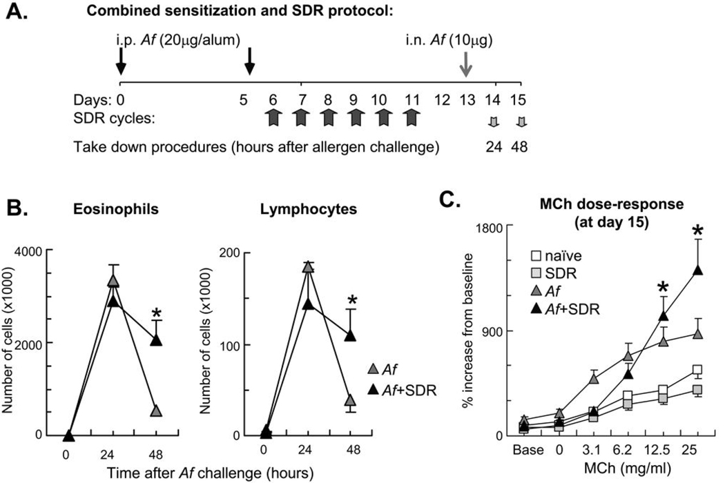
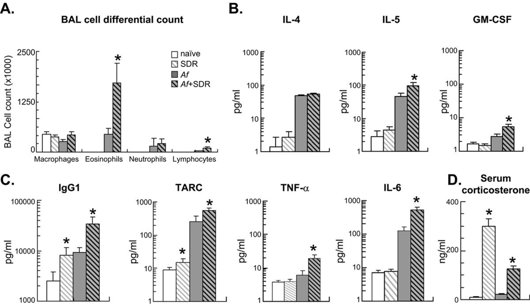
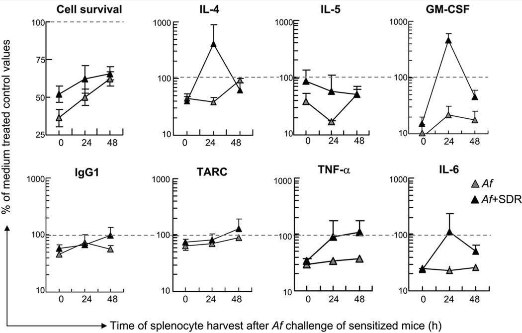
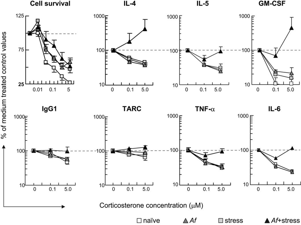
Chronic psychosocial stress exacerbates asthma, but the underlying mechanisms remain poorly understood. We hypothesized that psychosocial stress aggravates allergic airway inflammation by altering innate immune cell function. The effects of stress on airway inflammation, lung function, and glucocorticoid responsiveness were studied in a novel in vivo murine model of combined social disruption stress and allergic sensitization. The effects of corticosterone were assessed on cytokine profile and glucocorticoid receptor activation in LPS-stimulated spleen cell cultures in vitro. Airway inflammation resolved 48 h after a single allergen provocation in sensitized control mice, but not in animals that were repeatedly exposed to stress before allergen challenge. The enhanced eosinophilic airway inflammation 48 h after allergen challenge in these mice was associated with increased levels of IL-5, GM-CSF, IgG1, thymus-activated and regulatory chemokine, TNF-alpha, and IL-6 in the airways and a diminished inhibition of these mediators by corticosterone in LPS-stimulated splenocyte cultures in vitro. Stress-induced reduction of the corticosteroid effects paralleled increased p65 expression and a decreased DNA-binding capability of the glucocorticoid receptor in vitro. Furthermore, glucocorticoid receptor mRNA and protein expression in the lungs of mice exposed to both stress and allergen was markedly reduced in comparison with that in either condition alone or in naive mice. Thus, exposure to repeated social stress before allergen inhalation enhances and prolongs airway inflammation and alters corticosterone responsiveness. We speculate that these effects were mediated at least in part by impaired glucocorticoid receptor expression and function.


Figures

Figure 1
Figure 1
Combination of allergic airway sensitization and SDR caused delayed resolution of the inflammatory airway response and enhancement of allergic airway hyperresponsiveness at high methacholine concentrations. (A): In a combined protocol for allergic sensitization and SDR, mice received intraperitoneal (i.p.) Af in alum on days 0 and 5 followed by repeated SDR for 2 hours on days 6 through 11. Mice received intranasal (i.n.) allergen challenge on day 13 and were sacrificed either 24 or 48 hours later. Control naïve and SDR mice were sacrificed on day 15. (B): Combination of allergic airway sensitization and SDR increased number of airway eosinophils and lymphocytes 48h after Af challenge. BAL differential cell count was evaluated in Giemsa preparations. Naïve and SDR mice did not receive allergen challenge. There was no difference between naïve and SDR mice. (C): Exposure to allergen and stress enhanced airway hyperresponsiveness at high methacholine concentrations. Mice were studied 48 h after a single Af challenge. Mice received increasing doses of inhaled methacholine (MCh). Lung function was assessed using non-invasive plethysmography and the enhanced pause (Penh). There was no difference between naïve and SDR mice. Data are expressed as % changes from baseline. Baseline Penh measurements were as follows: naïve: 0.385±0.050; SDR:0.471±0.028; Af:0.924±0.367; Af+SDR:0.614±0.082 (B–C): Mean±SEM of n=5–8 each; *p<0.05 (Af vs. Af+SDR).
Figure 2
Figure 2
Mice were sensitized (i.p.) and challenged (i.n.) with Af extract and were also exposed to social stress (SDR) daily between days 7–12 as indicated. Sensitized mice were studied 48 h after a single Af challenge (on day 15). Naïve and SDR mice were also studied on day 15. (A): BAL differential cell count was evaluated in Giemsa preparations. Stress and allergic sensitization enhanced numbers of eosinophils and lymphocytes but not neutrophils and macrophages in the BAL fluid. There was no difference between naïve and SDR mice. (B–C): Chemokine, cytokine and immunoglobulin levels in the BAL were measured in the same groups by SearchLight technology. Increased eosinophilia was paralleled by enhanced levels of the eosinopoietic IL-5 and GM-CSF (B), increased levels of the B-cell derived IgG1 and the innate immune cell-derived TARC, TNF-α and IL-6 (C). (D): Stress significantly increased serum corticosterone levels p<0.01: SDR vs. naïve, Af or Af+SDR. Blood for endocrine measures was taken on day 15. (A–D): (Mean±SEM of n=8 per group) *p<0.05 Naïve vs. SDR and Af vs. Af+SDR; Repeated measure ANOVA followed by Tukey's HSD.
Figure 3
Figure 3
Combination of stress and allergen challenge attenuated the time-dependent inhibitory effect of corticosterone on splenocytes. Spleens were harvested before (0h), 24 and 48h after Af challenge. Splenocytes were cultured in the presence of LPS (1 µg/ml) and 5 µM of corticosterone for 48 hours. Cell viability (top left panel) was determined using a colorimetric assay. Cytokine, chemokine and immunoglobulin levels in the supernatant were measured by SearchLight technology. Data are presented as the percentage of cell viability or cytokine, chemokine and immunoglobulin concentration of control values measured in the absence of corticosterone, marked by dashed lines (Mean±SEM of n=5–8 per group).
Figure 4
Figure 4
Combination of stress and allergen challenge abolished the dose-dependent inhibitory effects of corticosterone on LPS-stimulated splenocytes. Spleens were harvested from naïve (open squares), or SDR controls (grey squares) and from Af−sensitized mice 24 h after Af challenge (Af, grey triangles, Af+SDR, black triangles). Splenocytes were cultured in the presence of LPS (1 µg/ml) and and 0, 0.01, 0.05, 0.1, 0.5 or 5 µM of corticosterone for 48 hours. Cell viability (top left panel) was determined using a colorimetric assay. Cytokine, chemokine and immunoglobulin levels in the supernatant were measured by SearchLight technology in each groups. Data are presented as the percentage of cell viability or cytokine, chemokine and immunoglobulin concentration of control values measured in the absence of corticosterone, marked by dashed lines (Mean±SEM of n=5–8 per group).
Figure 5
Figure 5
To study the effects of stress and asthma on function and expression of the glucocorticoid receptor (GR), splenocytes and whole lung tissue was investigated. Naïve mice (white bar) and mice subjected to stress (SDR, hatched bars), allergen (Af, grey bars) or allergen and stress (Af+SDR, grey hatched bars) were studied. On day 15 (48 h after Af challenge) spleens for GR nuclear translocation and lungs for GR mRNA and protein expression were harvested. (A): SDR inhibited nuclear translocation and GRE binding of the GR. Splenocytes were isolated and incubated in the presence of dexamethasone (10-7M). GR nuclear translocation was quantitated by an ELISA based TransAM GR kit with GRE-coated wells. Mean±STDEV of n=3 (duplicate experiments). Densitometric data are corrected for values obtained from HeLa cell positive controls. (B, D): Combination of Af and SDR reduced expression of GR protein. Total protein was isolated from spleens (B) and lung tissue (D). Western blot analysis was performed using an anti mouse GRα monoclonal antibody. Expression was assessed using densitometric analysis and GAPDH as control. Mean±SEM of n=6 (duplicate experiment). (C): Combination of SDR and Af reduced expression of GR mRNA in the lung tissue. Total RNA was isolated from lungs of mice. Reverse transcription and realtime PCR were performed. β2 macroglobulin mRNA was used as control. Data were analyzed using the ΔΔ Ct Method. Mean±SEM of n=4–6 (duplicate experiments) (A–D): *p<0.05 Af vs. Af+SDR

Similar articles

Cited by

References

    1. Isenberg SA, Lehrer PM, Hochron S. The effects of suggestion and emotional arousal on pulmonary function in asthma: a review and a hypothesis regarding vagal mediation. Psychosom. Med. 1992;54:192–216. - PubMed
    1. Lehrer PM, Isenberg S, Hochron SM. Asthma and emotion: a review. J. Asthma. 1993;30:5–21. - PubMed
    1. Beggs PJ, Curson PH. An integrated environmental asthma model. Arch. Environ. Health. 1995;50:87–94. - PubMed
    1. Heim C, Ehlert U, Hellhammer DH. The potential role of hypocortisolism in the pathophysiology of stress-related bodily disorders. Psychoneuroendocrinol. 2000;25:1–35. - PubMed
    1. Marshall GD, Jr, Agarwal SK. Stress, immune regulation, and immunity: applications for asthma. Allergy Asthma Proc. 2000;21:241–246. - PubMed

Publication types